Reading...
![]()
Play button
![]()
Play button
![]()
Use LEFT and RIGHT arrow keys to navigate between flashcards;
Use UP and DOWN arrow keys to flip the card;
H to show hint;
A reads text to speech;
25 Cards in this Set
- Front
- Back
- 3rd side (hint)
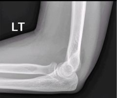
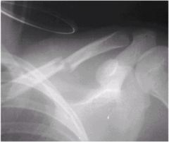
30 year old woman fell of a push-bike onto the left elbow. There is no open wound. She is neurovascularly intact
|
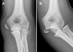
This is a LATERAL VIEW of an ELBOW of a SKELETALLY MATURE individual which displays an anterior and posterior sail sign with no definite fracture visible but is suggestive of a radial head fracture
|
|
|
Closed fracture
Neurovascularly intact |
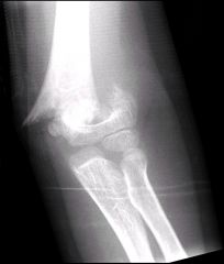
This is a PA view of an elbow of a skeletally immature individual which shows a complete transverse supracondylar fracture of the humerus with lateral displacement
|
Salter Harris Type I Classification
|
|
Closed fracture
Neurovascular intact |
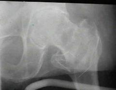
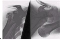
This is an AP view of the pelvis of a skeletally mature individual which shows a medially displaced intertrochanteric fracture
|
This is a stable fracture as it does not involve comminution of the posteromedial cortex
|
|

Closed
Neurovascularly Intact |
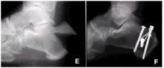
Lateral film of the ankle of a skeletally mature individual which shows:
Anterior process fracture (arrow 1) with associated talar neck fracture (arrow 2), and ankle instability (arrow 3). B. Postoperative reduction and fixation with screws, miniplate, and bone anchors. C. Healed fractures at 13 months. |
|
|

|
Extra-articular avulsion fracture A.
Lateral and axial x-rays. B. Initial CT scan sections showing nonarticular fracture. C. Further CT scan sections showing nondisplaced fracture into joint. D. Lateral and axial x-rays 1 year after simple cerclage band fixation |
|
|
|
E. Displaced avulsion fracture.
F. Five years after tension band with lag screw fixation. |
|
|

|
Medial process fracture of the calcaneus.
These vertical shear fractures are due to loading of the heel in valgus; they are seen on axial radiographs |
|
|
Closed Neurovascularly Intact
|
AP view of the clavicle of a skeletally mature individual which displays a transverse mid-shaft fracture of the clavicle with ~40% inferior displacement
|
|
|
Closed
Neurovascularly Intact |
AP view of the clavicle of a skeletally mature individual which displays an oblique extraarticular fracture of the distal clavicle
|
|
|
|
AP view of the humerus, skeletal maturity hard to determine. Highly comminuted fracture of the proximal humerus
|
|
|

Closed Neurovascularly Intact. What else do you want here?
|

This view was also necessary.
Just looking at the AP it appears as though this is just a simple one part fracture, but on the axillary it becomes apparent that it is a 2-part fracture with at the surgical neck. Formal reading: This is an AP and Axillary view of the shoulder of a skeletally mature individual. There is a transverse fracture with 100% displacement at the surgical neck of the humerus. This would be a Neer 2-part fracture |
|
|

Closed
Neurovascularly Intact |
AP view of the shoulder of a skeletally mature individual displaying a valgus-impacted 4-part fracture of the proximal humerus
|
|
|
Closed
Neurovascularly Intact |
This is an AP and Axillary view of the shoulder of a skeletally mature individual which displays a fracture at the lesser tuberosity and a barely-perceptible fracture at the surgical neck. This would be a Neer 2-part fracture
|
|
|

Closed
Neurovascularly intact |
This is an AP view of the left humerus and elbow of a skeletally mature individual which shows an oblique fracture of the midshaft of the humerus
|
|
|

Closed
Neurovascularly intact |

This is a lateral view of the elbow of a skeletally immature? individual which display a supracondylar humeral fracture with 100% displacement
This would be classified by Gartland and would be a Type III because it is completely displaced Closed reduction and pinning may be attempted |
|
|

Closed
Neurovascularly intact |
AP and lateral of the elbow of a skeletally mature individual which shows a transcondylar distal humerus fracture
If the fracture were open, unstable, or displaced, open reduction with plate fixation would be necessary Nonoperative treatment is indicated for nondisplaced or minimally displaced fractures |
|
|
Closed
Neurovascularly intact |
This is an AP and lateral of the elbow of a skeletally mature individual which displays an intercondylar distal humerus fracture
There appears to be some rotation of the medial epicondyle which would make this a Riseborough and Radin Type 3 |
|
|

Closed
Neurovascularly Intact |
This is an AP and lateral view of the elbow of a skeletally mature individual which displays a fracture of the medial condyle of the distal humerus.
This would be a Milch Type 1 injury |
|
|
Closed
Neurovascularly intact |

This is an AP and lateral of the elbow of a skeletally mature individual which shows a capitellar fracture of the distal humerus with extension into the trochlea.
Because the fracture line extends into the trochlea, it is classified as a Bryan and Morey Type IV fracture |
|
|

Closed
Neurovascularly Intact |

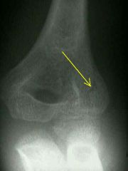
This is an AP and lateral of the elbow of a skeletally mature individual which displays a fracture of the capitellum of the distal humerus.
This would receive a Bryan and Morey Type 1 classification as there is a large osseous component of capitellum and there is no trochlear involvement |
|
|

Closed
Neurovascularly Intact |
This is an AP view of the elbow of a skeletally immature individual which displays a fracture of the lateral epicondyle
|
|
|

Closed
Neurovascularly Intact |

This is an AP view of the elbow of a skeletally immature individual which displays a fracture of the lateral epicondyle
|
|
|
Closed
Neurovascularly intact |

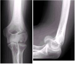
This is an AP and Lateral of the elbow of a skeletally mature individual which shows a displaced fracture of the medial epicondyle of the distal humerus
This could be treated with ORIF since the fractured piece is displaced, but if the piece cannot be reduced then it may be excised |
|
|

Closed
Neurovascularly Intact |

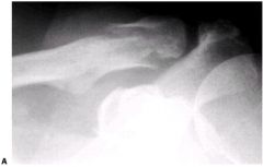
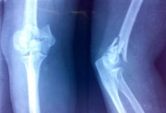
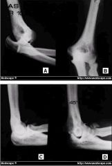
This is an AP and Lateral of the elbow of a skeletally mature individual which displays a displaced fracture of the radial head.
This would receive a Mason Type II classification as it involves a marginal fracture with displacement |
|
|
The first AP/Lateral is pre-reduction
The second AP/Lateral is post-reduction Closed Neurovascularly Intact |

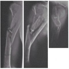
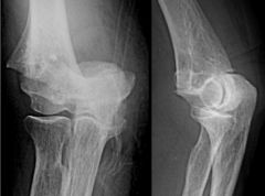
The first AP/Lateral shows a posterior dislocation of the elbow.
Post-reduction AP/Lateral makes apparent that there is a fracture of the radial head This would be classified as Mason Type IV because of the radial head fracture associated with dislocation of the elbow |
|