Reading...
![]()
Play button
![]()
Play button
![]()
Use LEFT and RIGHT arrow keys to navigate between flashcards;
Use UP and DOWN arrow keys to flip the card;
H to show hint;
A reads text to speech;
58 Cards in this Set
- Front
- Back
|
peritoneum has what two layers:
1. 2. and when and organ lies behind the peritoneum, it is said to be ... |
visceral
parietal retroperitoneal |
|
|
parietal peritoneum lies against what:
|
body wall
|
|
|
kidneys do not have a mesentary so they are ...
|
retroperitoneal
|
|
|
the kidneys are located in the ... wall around ... (vertebrae) and the right kidney is slightly (inferior/superior) to the left; why?
|
posterior abdominal
T12-L3 inferior because of the liver |
|
|
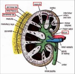
what are the 3 tissue layers surrounding the kidney:
1. 2. 3. |
1. Perirenal (perinephric) fat: on kidney
2. Renal fascia: external to fat 3. Pararenal (paranephric) fat: external to renal fascia |
|
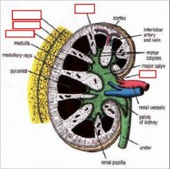
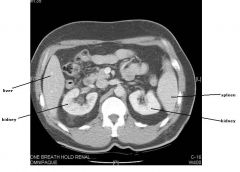
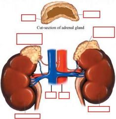
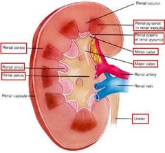
identify the labeled structures:
|
(see figure)
|
|
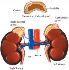
identify the labeled structure:
|
(see figure)
|
|

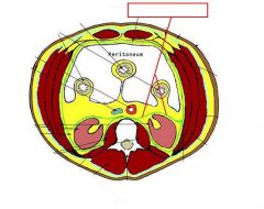
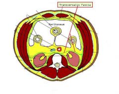
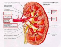
identify the labeled structures:
|
(see figure)
|
|
|
Renal hilum is ... leading into kidney and past the hilum is the ...
|
cleft; “doorway”
renal sinus |
|
|
... is the area at the center of the kidney where urine collects and is funneled into the ureter
|
renal pelvis
|
|
|
what is inside the renal sinus:
|
fat and vessels
|
|
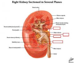
identify the labeled structures:
|

(see figure)
|
|
|
Structures entering or leaving renal hilum/sinus are:
1. 2. 3. |
1. renal vein
2. renal artery 3. renal pelvis |
|
|
where are the following structures located in the renal hilum:
1. renal vein 2. renal artery 3. renal pelvis |
1. renal vein - most anterior
2. renal artery - in middle 3. renal pelvis - most posterior |
|
|
what is contained inside the renal pelvis:
|
urine
|
|
|
what is the pathway of the urine:
|
minor calyces (many) --> major calyces (3-4) --> pelvis --> ureter --> urinary bladder
|
|
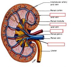
identify the labeled structures:
|
(see figure)
|
|
identify the labeled structures:
|
(see figure)
|
|
|
IVC is always to the ... and veins are ... (position)
|
right
anterior |
|
|
how many segmental branches are coming in to supply the kidneys:
|
5
|
|

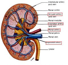
identify the labeled structures:
|
(see figure)
|
|
|
what is the vertebral levels for the renal arteries:
|
L1/L2
|
|
|
which renal artery is longer, right or left and why:
which renal vein is longer, right or left and why: |
right
the aorta sits to the left left ivc sits to the right |
|
|
right renal artery is (anterior/posterior) to IVC
|
posterior
|
|
|
on the left side only, the ... vein comes up to the left renal vein
(impt point) |
gonadal
|
|
|
where are the renal pyramids located:
|
in the medulla
|
|
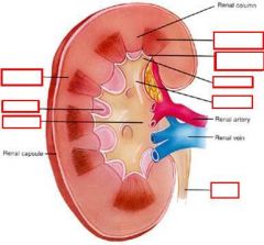
identify the labeled structures:
|
(see figure)
|
|
|
the outer layer of the kidney is the:
the inner layer is the: |
cortex
medulla |
|
|
the medulla consists of ... and ...
|
pyramids
renal columns |
|
|
where do the renal columns lie:
|
in between the pyramids
|
|
|
where are the papilla located:
|
apex of the pyramid
|
|
|
what covers the papilla of the pyramid:
|
minor calyx
|
|
|
what is the functional unit of the kidney:
|
nephron
|
|
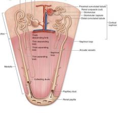
what is this structure
|

nephron - the functional unit of the kidney
|
|
|
where is the typical place to transplant a kidney:
|
pelvis
|
|
|
the ureter is ... long and is retroperitoneal and uses what type of muscle contraction to move the urine along
|
10 to 12 inches
peristalis |
|
|
what are the 3 common places for a kidney stone:
1. 2. 3. |
1. renal pelvis-ureter junction
2. pelvic brim (crosses over bone) 3. wall of urinary bladder |
|
|
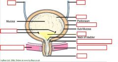
the bladder is (anterior/posterior) to pelvic organs and (anterior/posterior) to pubic symphysis and (is/is not) retroperitoneal
|
anterior
posterior is |
|
|
what is the muscle in the bladder wall called:
|
Detrusor muscle
|
|
|
... contraction acts as an active sphincter on the ureter while ... acts as a passive valve and both stops urine from going ...
|
detrusor
pressure of urine back up ureter |
|
|
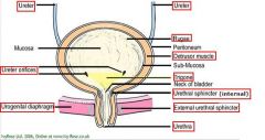
there is one triangular shaped area in the inside of the bladder that is smooth and is called the ... it is marked by:
|
trigone
2 ureters and the urethra |
|
|
there are 2 urethral sphincters
1. 2. |
1. External urethral sphincter 2. Internal urethral sphincter
|
|
|
1. External urethral sphincter (involuntary/voluntary)
2. Internal urethral sphincter (involuntary/voluntary) |
1. voluntary
2. involuntary |
|
identify the labeled structures:
|
(see figure)
|
|
|
what is the sympathetic innervation of the bladder:
parasympathetic: sensory (carried by parasympathetics): motor: somatic innervation: |
T11-L2
S2-S4 bladder distension detrusor muscle skeletal muscle of the sphincter urethrae |
|
|
where do the sympathetics go in the bladder:
|
sphincter vesicae
|
|
|
what is another name for:
1. Internal urethral sphincter 2. External urethral sphincter |
sphincter vesicae
sphincter urethrae |
|
|
adrenal glands are also called ... and press right up against the ... and are enclosed by ... and ...
|
suprarenal glands
diaphragm renal fascia perinephric fat |
|
|
for the adrenal glands there is a hilum for ... and ... but ... and ... enter at multiple sites
|
veins
lymphatics arteries nerves |
|
|
the adrenal cortex is derived from ... and produces 3 classes of steroid hormones:
1. 2. 3. |
mesoderm
1. glucocorticoids 2. mineralcorticoids 3. androgens |
|
|
the adrenal medulla is derived from ... and is part of the ... system but functions as ...
|
neural crest cells
nervous endocrine gland |
|
|
chromaffin cells are located where:
they are similar to ... and secrete |
medulla adrenal
postsynaptic, sympathetic neurons epinephrine & norephinephrine |
|
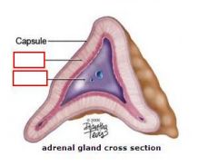
identify the labeled structures:
|
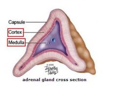
(see figure)
|
|
|
there are 3 different arteries coming to the adrenal gland, where do they go and from do they come:
1. 2. 3. |
1. Superior: from inferior phrenic a.
2. Middle: from aorta 3. Inferior: from renal a. |
|
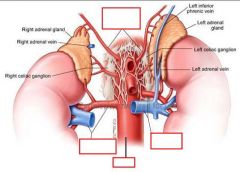
identify the labeled structures:
|
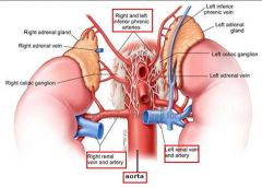
(see figure)
|
|
|
what hormone and gland control the adrenal cortex:
|
ACTH (adrenocorticotropic hormone)
pituitary gland |
|
|
what controls the adrenal medulla:
|
presynaptic fibers from greater
splanchnic nerve |
|
|
what is the neural pathway to the adrenal medulla:
|
presynaptic fibers from greater splanchnic nerve pass through celiac ganglia & synapse directly on cells of
adrenal medulla |