![]()
![]()
![]()
Use LEFT and RIGHT arrow keys to navigate between flashcards;
Use UP and DOWN arrow keys to flip the card;
H to show hint;
A reads text to speech;
15 Cards in this Set
- Front
- Back
|
What structures are associated with the following; anterior wall lateral wall posterior wall |
anterior wall: Sternum, costal cartilages, anterior intercostal spaces with their muscle conten lateral wall: ribs, intercostal spaces with their muscle content posterior wall: vertebral column, posterior ends of the ribs, posterior intercostal spaces with their muscle content |
|
|
Compare the external and internal inercostal muscles |
External intercostal muscles - Inspiratory muscles - Elevation of the ribs Internal intercostal muscles - Expiratory muscles - Depression of the ribs |
|
|
Compare the upper and lower rib margins of the intercostal spaces |
Upper rib margin & intercostal space: smaller vessels Lower rib margin: larger vessels |
|
|
Where are the upper and lower limits of the thoracic cavity? |
upper limit: thoracic inlet (T1-T4) lower limit: diaphragm |
|
|
List 6 structures in the thoracic inlet |
- Large aortic branches for head & neck - Large veins - Vagus and phrenic nerves - Oesophagus and Trachea - Sympathetic trunks - Apex of each lungs |
|
|
List 3 structures of the lower limit |
- central tendon - peripheral attachments - medial, lateral and intermediate crus |
|
|
What are the three openings in the diaphragm |
- inferior vena cava - oesophageal - aortic |
|
|
Which nerves supply the diaphragm? |
Phrenic nerve (cervical plexus: C3-C4-C5) |
|
|
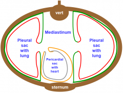
Draw and label the divisions within the thorax |
|
|
|
|
|
|
What is a hilum? |
organ inlet/outlet |
|
|
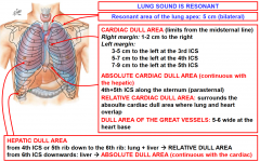
Draw a diagram identifying the following; - resonant area - cardiac dull area - absolute cardiac dull area - hepatic dull area |
|
|
|
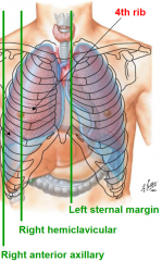
Where are the following margins located; left sternal margin right hemiclavicular margin right anterior axillary |
|
|
|
What are the two viscera that surround the heart? |
visceral pericardium: inner parietal pericardium: outer |
|
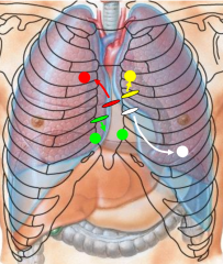
name the auscultation sites |
yellow: pulmonary red: aortic green: tricuspidal white: mitral |