![]()
![]()
![]()
Use LEFT and RIGHT arrow keys to navigate between flashcards;
Use UP and DOWN arrow keys to flip the card;
H to show hint;
A reads text to speech;
36 Cards in this Set
- Front
- Back
|
Classify the Temporomandibular joint |
Synovial, Biaxial, Condylar |
|
|
What movements are possible at the TMJ? |
- Elevation/Depression - Retraction/Protraction |
|
|
What are the articular surfaces of the TMJ? |
- Condyles of Mandible - Mandibular Fossa of Temporal bone |
|
|
What are the functions of the TMJ's articular disc? |
1. Improves articular fit 2. Divides joint into upper/lower cavities 3. Attachment site for lateral pterygoid |
|
|
Describe the attachment of the articular capsule in the TMJ |
- The articular capsule attaches to the mandibular fossa of the temporal bone, the neck of the mandible, and the articular disc - The capsule is TIGHT from the disc to the mandible and LOOSE from the disc to the fossa - This ensures the disc moves with the mandible |
|
|
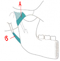
A - Lateral ligament of TMJ B - Stylomandibular ligament |
|
|
Where does the lateral ligament of the TMJ attach and what is its function? |
- Attaches from zygomatic bone to neck of mandible - Limits depression and retraction |
|
|
Where does the stylomandibular ligament attach and what is its function? |
- Attaches to styloid process of temporal bone to angle of mandible - limits depression and protraction |
|
|
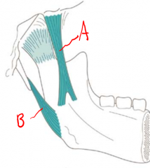
A - Sphenomandibular ligament B - Stylomandibular ligament |
|
|
Where does the sphenomandibular ligament attach and what is it's purpose? |
- The sphenomandibular ligament attaches to the sphenoid bone and the medial surface of the ramus of the mandible - limits depression and protraction |
|
|
Name the muscles involved in mastication |
1. Temporalis 2. Masseter 3. Lateral pterygoid 4. Medial pterygoid |
|
|
Which muscles are responsible for protraction of the mandible |
- Medial pterygoid - Lateral pterygoid - Masseter |
|
|
Which muscles are responsible for retraction of the mandible? |
Temporalis
|
|
|
What muscles are responsible for elevation of the mandible? |
- Masseter - Medial pterygoid - Temporalis |
|
|
What muscles are responsible for depression of the mandible? |
Depression is usually a gravity assisted motion, meaning the elevators are working ECCENTRICLY However, the lateral pterygoid may assist |
|
|
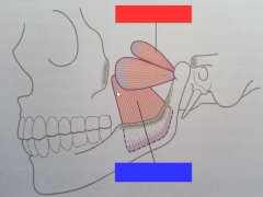
Red - Lateral Pterygoid Blue - Medial Pterygoid |
|
|
List the suprahyoid muscles |
1. Mylohyoid 2. Digastric 3. Stylohyoid |
|
|
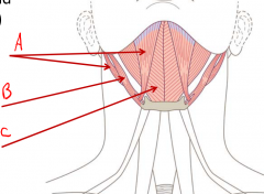
A - Digastric B - Stylohyoid C - Mylohyoid |
|
|
List the infrahyoid muscles |
1. Sternohyoid 2. Omohyoid 3. Thyrohyoid |
|
|
Which cranial nerve (name and number) supplies the muscles of mastication? |
V - Trigeminal |
|
|
Which cranial nerve (name and number) supplies the muscles of facial expression? |
VII - Facial |
|
|
Which cranial nerve (name and number) supplies the skin of the face? |
V - Trigeminal |
|
|
What nerve supplies the infrahyoid muscles? |
Cervical plexus |
|
|
What nerve supplies the suprahyoid muscles? |
- Mylohyoid and anterior belly of digastric supplied by Trigeminal nerve - Stylohyoid and posterior belly of digastric supplied by Facial nerve |
|
|
Which nerve supplies the lining of the mouth and nose? |
Trigeminal |
|
|
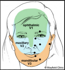
How is the sensory innervation of the face distributed? |
|
|
|
What are the 3 sensory divisions of the trigeminal nerve? |
1. Opthalmic 2. Maxillary 3. Mandibular |
|
|
Which nerve supplies skin of the neck? |
Cervical plexus |
|
|
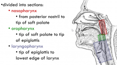
Name the divisions of the pharynx and list the boundaries of each division |
|
|
|
A) In the external muscle layer of the pharynx, how are the muscles oriented and what is their purpose? B) What nerve innervates these muscles? |
A - The muscles are oriented in a circular manner to constrict the pharynx B - The Vagus Nerve (X) |
|
|
A) In the internal muscle layer of the pharynx, how are the muscles oriented and what is their purposed? B) What nerve innervates these muscles? |
A - The muscles are oriented ina a longitudinal manner to elevate the pharynx B - Vagus (X) and Glossopharyngeal (IX) |
|
|
Which nerve is responsible for sensory innervation of the pharynx? |
Vagus X |
|
|
Briefly describe the first stage of swallowing |
Food is voluntarily moved from the oral cavity to the pharynx |
|
|
Briefly describe the second stage of swallowing |
Food involuntarily moves through pharynx to the oesophagus |
|
|
Describe the muscle activity that occurs in the first stage of swallowing and describe the role of each |
- Buccinators prevent the food from moving laterally to the teeth - Suprahyoid muscles raise the floor of the mouth - Intrinsic tongue muscles raise tip of tongue to hard palate - Extrinsic tongue muscles raise sides of tongue to create a chute |
|
|
Describe the muscle activity that occurs in the second stage of swallowing and describe the roles of each |
- Soft palate is raised to close opening to nasopharynx. (stop food entering nose) - Internal pharyngeal muscles elevate the pharynx - Suprahyoid muscles raise the hyoid and close the laryngeal inlet. (stop food entering airway) - Tongue bulges over opening of oropharynx (stop food reentering oral cavity) - Pharyngeal muscles (int + ext) work together in peristalsis to move food through pharynx |