![]()
![]()
![]()
Use LEFT and RIGHT arrow keys to navigate between flashcards;
Use UP and DOWN arrow keys to flip the card;
H to show hint;
A reads text to speech;
22 Cards in this Set
- Front
- Back
|
What are the medial, lateral, superior boundaries of the femoral triangle? Which structures make the floor and roof of the femoral triangle? |
M-medial border of adductor longus L-medial border of Sartorius Superior- inguinal ligament Floor- adductor longus, pectineus, psoas and iliacus Roof- superficial fascia containing superficial LNs, great saphenous vein and its tributaries, deep fascia i.e. fascia lata which is pierced by the great saphenous vein |
|
|
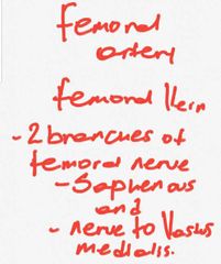
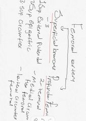
What are the contents of the femoral triangle? |
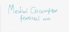
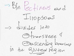
Femoral sheath Femoral artery and it's branches Femoral vein and its tributaries Femoral nerve and its branches Deep inguinal LNs Profunda femoris artery and veins and their circumflex branches (lateral and medial) |
|
|
Femoral sheath origin and contents |
Wall of the sheath is derived from extraperitoneal fascia I.e. transversalis fascia anteriorly and iliaus fascia posteriorly
3 compartments and 2 septa |
|
|
The 3 compartments of the femoral sheath |
Lateral compartment Middle compartment and Medial compartment |
|
|
Lateral compartment of femoral sheath |
Femoral artery and genital branch of genitofemoral nerve |
|
|
Middle compartment |
Femoral vein |
|
|
Medial compartment- Femoral canal |
Lymph vessels, deep LNs and fat Site of femoral hernia 0.5 inch admits the tip of the little finger Dead space for expansion of the distended femoral vein Lymphatic pathway from LL to the external iliac nodes |
|
|
Boundaries of the femoral canal Anteriorly Medially Posteriorly and Laterally |
A- Inguinal ligament M- lacunar ligament L-femoral vein P- pectineal ligament |
|
|
Lacunar ligament |
Pectineal part of inguinal ligament From the inguinal ligament and pubic tubercle to pectineal line |
|
|
Pectineal ligament |
The thickened periosteum along the pectineal border of the superior pubic ramus Medial continuation of lacunar ligament |
|
|
Femoral hernia |
Sac may emerge through the saphenous oppening then turns upward along the pathway of superficial circumflex iliac and superficial epigastric vessels Project above the inguinal ligament |
|
|
Cannal of hunter / subsartorial canal |
Adductor canal From the apex of the femoral triangle Ends 10cm above the adductor tubercle |
|
|
Boundaries of the adductor canal Posteriorly Anterolaterally Anteromedially |
P-Adductor longus and Magnus AL- vastus medialis AM- Sartorius Sartorius forms the roof |
|
|
|
|
|
|
|
|
|
|
|
|
|
|
|
|
|
|
|
|
|
|
|
|
|
|
|