![]()
![]()
![]()
Use LEFT and RIGHT arrow keys to navigate between flashcards;
Use UP and DOWN arrow keys to flip the card;
H to show hint;
A reads text to speech;
110 Cards in this Set
- Front
- Back
- 3rd side (hint)
|
Basic structural unit of fungi |
Hypha |
|
|
|
Fungi are also classified as |
Thallophytes |
|
|
|
Heterotropic members of the plant family that lack stems and roots |
Fungi |
|
|
|
Multicellular phase, cottony, mycelial mass |
Mold phase |
|
|
|
Unicellular phase, creamy, resembling bacterial colony |
Yeast phase |
|
|
|
Fungi that stays in mold phase at 25 & 37 degrees celcius |
Monomorphic |
|
|
|
Fungi in mold phase at 25 & 37 degrees celcius, and yeast phase in tissue |
Diphasic |
|
|
|
Fungi in mold phase at 25 degrees celcius and becomes yeast at 37 degrees celcius or when in tissue |
Dimorphic |
|
|
|
An intertwining structure composed of hyphae |
Mycelium |
|
|
|
Hyphae having no cross-walls or divisions |
Aseptate hyphae |
Also known as coenocytic hyphae |
|
|
Hyphae having cross-walls or divisions |
Septate hyphae |
|
|
|
Vegetative portion of the mycelium |
Thallus |
|
|
|
Reproductive part of the mycelium containing the fruiting bodies |
Aerial |
|
|
|
Spores enclosed in ascus |
Ascospores |
|
|
|
Spores formed by the fusion of two identical cells from the same hypha |
Zygospores |
|
|
|
Spores formed by the fusion of two identical cells from different hyphae |
Oospores |
|
|
|
Spores enclosed in club-shaped basidium |
Basidiospores |
|
|
|
Spores arising from the side of hyphae |
Conidia |
|
|
|
Small, unicellular conidia |
Microconidia |
|
|
|
Large, multicellular conidia |
Macroconidia |
|
|
|
Conidia from simple budding |
Bastoconidia |
|
|
|
Thick-walled spores formed during unfavorable environmental conditions |
Chlamydoconidia |
|
|
|
3 types of chlamydoconidia |
Terminal Intercalary Sessile |
|
|
|
Formed from the fragmentation of mycelium; rectangular or barrel shaped |
Arthroconidia |
|
|
|
Mycosis in which there is no cellular response by the host |
Superficial mycoses |
|
|
|
Uneven pigmentation of the skin caused by M furfur |
Ptyriasis or Tinea vesicolor |
|
|
|
Morphology of M furfur |
"Spaghetti and meatballs" |
|
|
|
Brown-black cross outside hairshaft |
Black piedra |
|
|
|
Causes black piedra |
Piedraia hostae |
|
|
|
Light brown nodules on beard |
White piedra |
|
|
|
Causes white piedra |
Trichosporon beigelii |
|
|
|
Also known as dermatomycoses |
Cutaneous mycoses |
|
|
|
3 keratinized tissues |
Skin Hair Nails |
|
|
|
Caused by dermatophytes |
Tinea or ringworm |
|
|
|
Anthrophilic microsporum (infects man) |
Microsporum audouinii |
|
|
|
Colonizes skin and hair |
Microsporum |
|
|
|
Tinea capitis is seen in |
Scalp |
|
|
|
Tinea barbae is seen in |
Beard |
|
|
|
Tinea corpuris is seen in |
Body |
|
|
|
Tinea cruris is seen in |
Groin |
|
|
|
Infection of the groin caused by Tinea cruris |
Jock's itch |
|
|
|
Tinea ungmium is seen in |
Nails |
|
|
|
Tinea pedis is seen in |
Feet |
|
|
|
Manifestation of Tinea pedis |
Athlete's foot |
|
|
|
Dermatophyte having no microconidia |
Epidermophyton |
|
|
|
Dermatophyte which colonizes skin, hair and nails |
Trichophyton |
|
|
|
Indicator for Dermatophyte test medium (DTM) |
Phenol red |
|
|
|
Large spindle shaped rough macroconidia, curved terminal ends, and positive for rice medium |
M canis |
|
|
|
M canis produce what color fluorescence of ectothrix hairs |
Green-yellow |
|
|
|
M audouinii produce what color fluorescence of ectothrix hairs |
Apple green |
|
|
|
Pencil shaped macroconidia, tear shaped microconidia, urease negative, hair baiting negative |
T rubrum |
|
|
|
V-shaped penetration of hairshaft, spherical grape-like clusters-microconidia |
T mentagrophytes |
|
|
"Balloon" form |
T tonsurans |
|
|
"Rat tail" macroconidia |
T verrucosum |
|
|
Favus type of Tinea capitis, "favic chandeliers" |
T schoenleinii |
|
|
|
Swollen hyphae containing cytoplasmic granules |
T violaceum |
|
|
|
Dark, slow-growing fungi |
Dematiaceous fungi |
|
|
|
Also known as mixed sporulation |
Fonsecaea |
|
|
|
Conidia in side |
Acrotheca |
|
|
|
Conidia in chain |
Cladosporium |
|
|
|
Conidia in cluster |
Phialophora |
|
|
|
Granulomatous tumor of subcutaneous tissue |
Mycetoma |
|
|
|
2 fungus-like bacteria |
Nocardia Actinomyces |
|
|
|
Most common cause of mycetoma |
Pseudoallescheria boydii |
|
|
|
Rare condition caused by dematiaceous saprobes |
Phaeohyphomycosis |
|
|
|
"Rose garden's disease" is also known as |
Sporotrichosis |
|
|
|
Causes sporotrichosis |
Sporothrix schenckii |
|
|
|
Appearance of S schenckii conidia in room temperature |
"Flowerette" conidia |
|
|
|
Concentric radiating eosinophilic material caused by antigen-antibody reaction |
Asteroid bodies |
|
|
|
Also known as Gilchrist's disease |
North American Blastomycosis |
|
|
|
Causative agent of North American Blastomycosis |
Blastomyces dermatitidis |
|
|
|
Appearance of B dermatitidis conidia in room temperature |
"Lollipop" conidia |
|
|
|
Exoantigen test for B dermatitidis |
Specific A band |
|
|
|
Causative agent of South American Blastomycosis |
Paracoccidioides brasiliensis |
|
|
|
Causative agent of Darling's disease |
Histoplasma capsulatum |
|
|
|
Tuberculate macroconidia, no microconidia, monomorphic fungi |
Sepedonium |
|
|
|
Exoantigen test for Histoplasma capsulatum |
H and/or M bands |
|
|
|
Also known as desert fever |
San Joaquin Valley fever |
|
|
|
Causative agent of san Joaquin Valley fever |
Coccidioides immitis |
|
|
|
Major biological hazard to lab personel |
Coccidioides immitis |
|
|
|
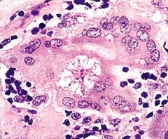
Appearance of Coccidioides immitis at 37 degrees celcius |
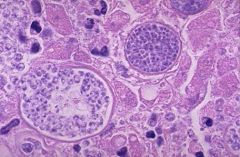
Spherule filled with endospores |
|
|
|
Causative agent of trush and vulvovaginitis, positive for germ tube formation |
Candida albicans |
|
|
|
Causative agent of torulosis or torulopsis, appear as encapsulated yeast cell in bird and bat droppings |
Cryptococcus neoformans |
|
|
|
Also known as nigrosine stain |
India ink stain |
|
|
|
Test for cryptococcal antigen in CSF |
Latex agglutination test |
|
|
|
Used to dissolve keratin |
10% KOH mount |
|
|
|
Color of positive hyphae in Periodic Acid Schiff (PAS) |
Purplish-red |
|
|
|
Gram stain modification used for fungi |
Hucker modification |
|
|
|
All fungi are gram |
Positive |
|
|
|
Culture for fungi with acidic pH as inhibitor for bacteria |
Sabouraud dextrose agar (SDA) |
|
|
|
Mycosel or Mycobiotic contains what 2 inhibitors |
Chloramphenicol (for bacteria) Cycloheximide (for saprophytic fungi) |
|
|
|
Culture media used to differentiate T rubrum from T metagrophytes |
Cornmeal agar |
|
|
|
Culture media for aspergilus |
Czapek's agar |
|
|
|
Aspergilus causes |
"Farmer's lungs" |
|
|
|
C neoformans on Staib's medium produces what color of colonies |
Brown-black colonies |
|
|
|
Staib's medium is also known as |
Birdseed or Nigerseed medium |
|
|
|
Cottonseed medium is used for |
B dermatitidis |
|
|
|
Positive for rice medium |
M canis |
|
|
|
Incubation temperature of fungal cultures |
25-30 degrees celcius (room temperature) Optimum at 30 degrees celcius |
|
|
|
Fungal cultures are held for how many days |
30 days |
|
|
|
Positive for L-DOPA ferric citrate test |
Cryptococcus neoformans |
|
|
|
QC for urease test |
C neoformans (positive) C albicans (negative) |
|
|
|
2 organisms that produce red colonies on SDA |
T rubrum T mentagrophytes |
|
|
|
QC for hair baiting test |
T mentagrophytes T rubrum |
|
|
|
Serologic confirmation test for systemic fungi |
Exoantigen test |
|
|
|
Biosafety level of organisms not harmful to healthy individuals |
Biosafety level 1 |
|
|
|
Biosafety level of common agents of infectious disease including HIV |
Biosafety level 2 |
|
|
|
Organisms classified as Biosafety level 3 |
MTB Systemic fungi Organisms grown in large quantities F tularensis Brucella species |
|
|
|
Viruses classified as Biosafety level 4 |
Arenavirus Arbovirus Filovirus (Ebola) Smallpox |
|
|
|
What year was smallpox eradicated from the world? |
1979 |
|