![]()
![]()
![]()
Use LEFT and RIGHT arrow keys to navigate between flashcards;
Use UP and DOWN arrow keys to flip the card;
H to show hint;
A reads text to speech;
38 Cards in this Set
- Front
- Back

|
Circumoral glands in the cat - large sebaceous glands - more numerous in lower lip - secretions used for scent marking |
|
Muscles of the head |
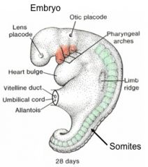
Somatic mm. develop from somites - extrinsic eye mm., tongue mm.
Visceral mm. develop from pharyngeal arches - mm. of mastication (1st PA) - mm. of facial expression (2nd PA) - pharyngeal constrictors (3rd PA) - mm. of pharynx, larynx, esophagus (4th PA) - COST mm., laryngeal mm. (5th & 6th PA) |
|
|
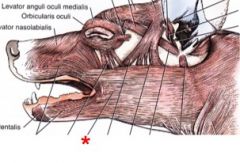
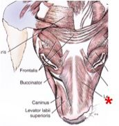
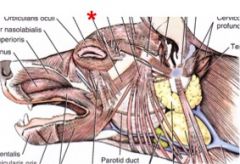
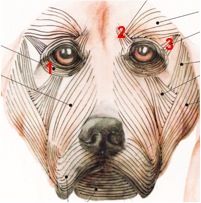
Facial muscles |
Involved with facial expression - different from mm. of mastication (which raise/lower mandible to open/close mouth) - innervated by CN VII (facial n.) |
|
|
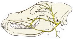
1) Facial n.(CN VII) - exits skull caudal to acoustic meatus through stylomastoid foramen - brs. into: 2) auriculopalpebral n. 3) dorsal buccal br. 4) ventral buccal br. 5) cervical br.
|
|
|
Platysma m. (a cutaneous m.) O: dorsal median raphe of neck I: angle of the mouth; radiates into orbicularis oris m. N: dorsal & ventral buccal brs. & caudal auricular br. of facial n. A: draw commissure of lips caudally |
|

|
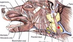
Orbicularis oris m. - located along free border of lips around angle of mouth N: dorsal & ventral buccal brs. of facial n. A: close mouth, pull nose downward during sniffing, dilate nostril |
|

|
Buccinator m. - forms foundation of cheek (deep to platysma and partly deep to orbicularis oris) I: alveolar margins of mandible & maxilla, & buccal mucosa N: dorsal and ventral buccal brs. of facial n. A: move food from vestibule to masticatory surface of teeth |
|
|
Levator nasolabialis m. O: frontal region btwn orbits & maxillary bone I: edge of upper lip & external naris (nostril) N: auriculopalpebral br. of facial n. A: dilate nostril, raise upper lip |
|
|
Zygomaticus m. O: rostral angle of scutiform cartilage I: commissure of lip N: auriculopalpebral br. of facial n. A: draw commissure of mouth dorsocaudally; draw scutiform cartilage (and ear) rostrally |
|
|
Parotidoauricularis m. O: caudal to laryngeal region, on or near ventral midline (blends with cervical fascia) I: antitragus of ear N: facial n. A: depress the ear |
|
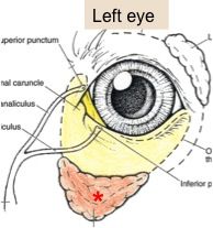
Eyelids |
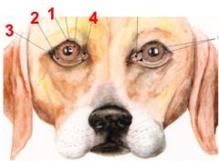
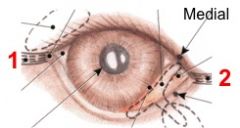
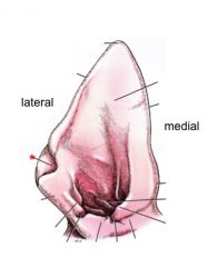
Palpebra (pl - palpebrae) 1) superior eyelid 2) inferior eyelid Palpebral fissure - opening btwn palpebrae Commissures (canthi) = corners 3) lateral palpebral commissure 4) medial palpebral commissure |
|
Eyelids |
Lacrimal caruncle - triangular prominence at medial angle of eye - has small, fine hairs & sebaceous glands |
|
Eyelids |
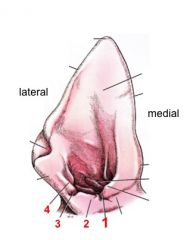
2) Medial palpebral ligament - attaches medial palpebral commissure to frontal bone 1) Lateral palpebral ligament - attaches lateral palpebral commissure to zygomatic bone Eyelashes - present on superior palpebra, absent on inferior palpebra |
|
|
Tear film: Superficial layer |
- Lipids secreted by tarsal (Meibomian) and ciliary glands located in palpebrae - Limits evaporation of underlying aqueous layer - Provides binding effect for tear film b/c of high surface tension |
|
|
Tear film: Middle aqueous layer |
- Watery substance from lacrimal gland & superficial gland of 3rd eyelid - Flushes foreign material from conjunctival sac & lubricates palpebral/corneal interfaces |
|
|
Tear film: Inner mucoid layer |
-Mucoproteinaceous product of conjunctival cells -Located btwn aqueous layer and cornea -Binds aqueous film to cornea |
|
|
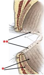
*Tarsal (Meibomian) glands -modified sebaceous glands -secrete lipids, part of superficial layer of tear film -better developed in superior eyelid **Ducts open at eyelid margin |
|
|
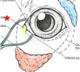
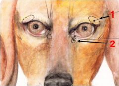
1) Lacrimal gland - dorsolateral to eye 2) Superficial gland of 3rd eyelid - ventromedial to eye
-both produce aqueous portion of tears -innervated by facial n. (parasympathetic) |
|
|
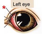
Third eyelid -aka plica semilunaris or nictitating membrane -comprised of a fold of palpebral conjunctiva and a T-shaped piece of cartilage -lymphoid tissue located on medial surface |
|
|
Superficial gland of the 3rd eyelid -surrounds ventral portion of 3rd eyelid |
|
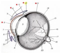
Conjunctiva |
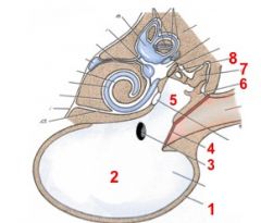
Mucous membrane -Produces inner mucoid layer of tear film -Palpebral conjunctiva (10) covers inner surface of palpebraa -Bulbar conjunctiva (11) covers rostral part of sclera, continuous with outer layer of cornea (8) -Conjunctiva fornix (12) junction of palpebral & bulbar conjunctiva -Conjunctival sac is space btwn eyeball & palpebra |
|
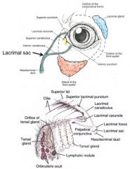
Aqueous tear flow |
-Lacrimal gland > thru ducts in superior palpebra >over anterior surface of eyeball > conjunctival sac located ventrally -through superior & inferior lacrimal ducts (canaliculi) -through lacrimal sac w/in lacrimal fossa of lacrimal bone -through nasolacrimal duct > nasal vestibule |
|
|
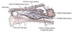
Nasolacrimal duct -begins at lacrimal sac, located in lacrimal fossa of lacrimal bone -courses through lacrimal canal of lacrimal & maxillary bones -rostral to conchal crest, courses deep to nasal mucosa, along side of maxilla -ends in nasal vestibule on ventral aspect of alar fold |
|
Eyelid muscles |
1) Orbicularis oculi m. - encircles the eye, closes palpebral fissure 2) Levator anguli oculi medialis m. - dorsomedial to eye, raises the eyelid 3) Retractor anguli oculi lateralis m. - covers lateral palpebral ligament - retracts lateral angle of eye caudally (squint) Innervation: palpebral brs. of auriculopalpebral br. of facial n. (CN VII) |
|
|
Extrinsic ear mm. |
3 groups: -Rostral auricular mm. -Dorsal auricular mm. -Caudal auricular mm. |
|
External ear |
1) auricle (aka pinna) 2) ear canal (external acoustic meatus) - 2 parts: vertical & horizontal canals |
|
|
External ear: -auricular cartilage |
-funnel shaped -forms vertical ear canal, part of horizontal ear canal |
|
|
External ear: -annular cartilage |
-btwn auricular cartilage and external acoustic meatus of temporal bone -forms part of horizontal ear canal |
|
|
External ear: -scutiform cartilage |
-small plate in muscles rostral and medial to the ear |
|
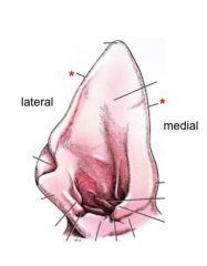
External ear (right) |
Helix -lateral and medial borders |
|
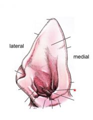
External ear (right) |
Anthelix -transverse ridge on inner aspect of auricle |
|
External ear (right) |
Marginal cutaneous sac -aka cutaneous marginal pouch -skin pouch |
|
External ear (right) |
1) Pretragic incisure 2) Tragus 3) Intertragic incisure 4) Antitragus |
|
|
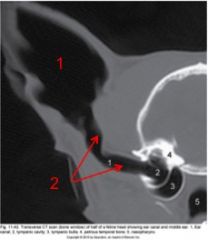
Middle ear - air filled 1) tympanic bulla 2) tympanic cavity 3) Internal auditory (Eustachian) tube-connects to nasopharynx 4) tympanic membrane (ear drum) 5) epitympanic recess 6) malleus 7) incus 8) stapes |
|
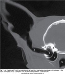
Middle ear - Cat |
Septum bullae divides tympanic bulla into two compartments |
|
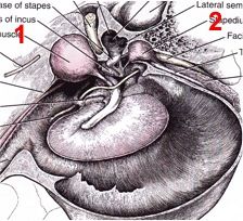
Middle ear mm. |
Tensor tympani m. -attaches to malleus
Stapedius m. -attaches to stapes -smallest m. in the body |
|
|
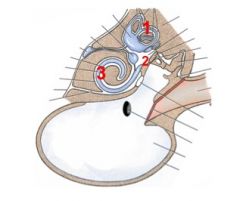
Inner ear - in petrous temporal bone -Vestibular system: 1) semicircular canals 2) vestibule -Hearing 3) cochlea |
|
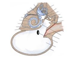
Inner ear fluids |
Perilymph - fills white spaces
Endolymph - fills blue spaces |