Reading...
![]()
Play button
![]()
Play button
![]()
Use LEFT and RIGHT arrow keys to navigate between flashcards;
Use UP and DOWN arrow keys to flip the card;
H to show hint;
A reads text to speech;
99 Cards in this Set
- Front
- Back
|
What is hematopoiesis / hemopoiesis?
|
Formation and development of various types of blood cells and other formed elements
|
|
|
Where does hematopoiesis take place in the adult?
|
Red Bone Marrow present in skull, vertebrae, ribs, sternum, ilia, and proximal epiphyses of some long bones
|
|
|
What replaces the red bone marrow in bony cavities where hematopoiesis does not take place?
|
Yellow Bone Marrow (infiltrated w/ fat)
|
|
|
What does hematopoiesis begin with?
|
Pluripotent hematopoietic stem cells (HSCs)
|
|
|
What do the progenitor cells from HSCs develop?
|
Colony Forming Units (CFUs) that generate a given type of blood cell
|
|
|
By what process are RBCs formed?
|
Erythropoiesis
|
|
|
What are the goals of Erythropoiesis?
|
Form a cell which is:
- Small to circulate easily - Contains abundant hemoglobin for gas exchange - Biconcave to maximize surface area for gas exchange |
|
|
What happens to the color of the cytoplasm in Erythropoiesis?
|
Change from Blue (polyribosomes, Hb mRNAs) to Red (protein, Hemoglobin)
|
|
|
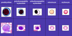
What are the precursors of RBCs during Erythropoiesis that we should be able to identify?
|

1. Proerythroblast
2. Polychromatophilic Erythroblast 3. Orthochromatophilic Erythroblast 4. Reticulocyte 5. Erythrocyte |
|
|
Proerythroblast:
- Stage - Cell size - Nucleus - Cytoplasm |
- Stage: 1st
- Cell size: large (>2x size of RBC) - Nucleus: large (~80% cell volume), round, central, prominent nucleolus - Cytoplasm: basophilic (dark/royal blue) d/t presence of polyribosomes (Hb mRNA) |
|
|
Polychromatophilic Erythroblast:
- Stage - Cell size - Nucleus - Cytoplasm |

- Stage: 2nd
- Cell size: medium (1.5x size of RBC) - Nucleus: small, round, condensed chromatin - Cytoplasm: grayish blue |
|
|
Orthochromatophilic Erythoblast:
- Stage - Cell size - Nucleus - Cytoplasm |

- Stage: 3rd
- Cell size: small (slightly larger than mature RBC) - Nucleus: small, round, eccentrically located, very condensed chromatin - Cytoplasm: cytoplasm staining identical to that of mature RBC |
|
|
Retiuclocyte:
- Stage - Cell size - Nucleus - Cytoplasm |

- Stage: 4th
- Cell size: cannot be distinguished from mature RBC - Nucleus: cannot be distinguished from mature RBC - Cytoplasm: cannot be distinguished from mature RBC **Distinguishable from RBCs when stained w/ a supravital dye (eg, cresyl blue) because residual polyribosomes stain blue ** |
|

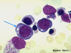
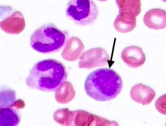
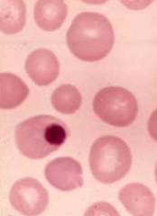
What kind of cell is this? How can you tell?
|

Proerythroblast
- Stage: 1st - Cell size: large (>2x size of RBC) - Nucleus: large (~80% cell volume), round, central, prominent nucleolus - Cytoplasm: basophilic (dark/royal blue) d/t presence of polyribosomes (Hb mRNA) |
|
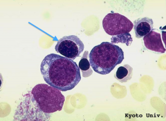
What kind of cell is this? How can you tell?
|

Polychromatophilic Erythroblast
- Stage: 2nd - Cell size: medium (1.5x size of RBC) - Nucleus: small, round, condensed chromatin - Cytoplasm: grayish blue |
|

What kind of cell is this? How can you tell?
|
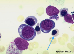
Orthochromatophilic Erythroblast
- Stage: 3rd - Cell size: small (slightly larger than mature RBC) - Nucleus: small, round, eccentrically located, very condensed chromatin - Cytoplasm: cytoplasm staining identical to that of mature RBC |
|
What kind of cell is this? How can you tell?
|

Reticulocyte:
- Distinguishable from RBCs when stained w/ a supravital dye (eg, cresyl blue) because residual polyribosomes stain blue |
|
|
How do you distinguish a reticulocyte from a fully mature erythrocyte?
|

- Stain w/ a supravital dye (eg, cresyl blue)
- Residual polyribosomes stain blue |
|
|
What product is being synthesized by polyribosomes in cells in early stages of erythrocyte production?
|
Hemoglobin
|
|
|
Why is the cytoplasm changing color as the cells in the erythropoietic series differentiate?
|

- Increase in Hemoglobin protein content leads to increased red staining
- Decrease in polyribosome (Hb mRNA) content leads to decreased blue staining |
|
|
How might iron deficiency affect morphology of erythrocytes?
|
- Results in RBCs which are microcytic (smaller than normal)
- Exhibit hypochromasia (less Hb leads to decreased redness in RBCs) - They tend to have staining only at periphery of cell d/t increased central pallor |
|
|
At which stage of development do the cells in the erythropoietic series lose their miotic ability?
|
Early normoblasts may divide, but reticulocytes which lack nuclei are obviously post-mitotic
|
|
|
If reticulocytes are found in the peripheral blood, does that indicate a pathological condition?
|
- No, reticulocytes normally complete their maturation during the first 24-48 hours in circulation
- Excessive reticulocytes in peripheral blood is indicative of certain anemias especially hemolytic anemia, subacute hemorrhage, or ascent to high altitude |
|
|
Erythropoietin is a glycoprotein hormone that promotes development of erythroid progenitor cells by inhibiting programmed cell death. What tissue normally produces erythropoietin?
|
Kidney (and liver)
|
|
|
What are the precursors of neutrophils during Granulopoiesis that we should be able to identify?
|
1. Myeloblast
2. Promyelocyte 3. Myelocyte 4. Metamyelocyte 5. Band / Stab Cell 6. Segmented Neutrophil |
|
|
Myeloblast:
- Stage - Cell size - Nucleus - Cytoplasm |

- Stage: 1
- Cell size: large - Nucleus: large, round, occupies the majority of the cell volume, delicate chromatin, prominent nucleoli - Cytoplasm: thin rim of light blue cytoplasm |
|
|
Promyelocyte:
- Stage - Cell size - Nucleus - Cytoplasm |

- Stage: 2
- Cell size: large, larger than a myeloblast - Nucleus: large, round, delicate chromatin, prominent nucleoli - Cytoplasm: abundant, heavily granulated by primary granules which obscure the nucleus |
|
|
Myelocyte:
- Stage - Cell size - Nucleus - Cytoplasm |

- Stage: 3
- Cell size: large - Nucleus: not indented - Cytoplasm: specific granules present |
|
|
Metamyelocyte:
- Stage - Cell size - Nucleus - Cytoplasm |

- Stage: 4
- Cell size: smaller than myelocytes - Nucleus: kidney-shaped / indented (<1/2 of diameter) - Cytoplasm: pink granules |
|
|
Band / Stab Cell:
- Stage - Cell size - Nucleus - Cytoplasm |
- Stage: 5
- Cell size: approximately same as mature cell - Nucleus: horseshoe-shaped / deeply indented (>1/2 of diameter) - Cytoplasm: pink granules |
|
|
Mature Neutrophil:
- Stage - Cell size - Nucleus - Cytoplasm |

- Stage: 6
- Cell size: mature size - Nucleus: multiple nuclear lobes (3-5) separated by thin nuclear filament, called a "segment" - Cytoplasm: pink granules |
|

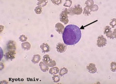
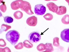
What kind of cell is this? How can you tell?
|
Myeloblast
- Stage: 1 - Cell size: large - Nucleus: large, round, occupies the majority of the cell volume, delicate chromatin, prominent nucleoli - Cytoplasm: thin rim of light blue cytoplasm |
|
What kind of cell is this? How can you tell?
|

Promyelocyte
- Stage: 2 - Cell size: large, larger than a myeloblast - Nucleus: large, round, delicate chromatin, prominent nucleoli - Cytoplasm: abundant, heavily granulated by primary granules which obscure the nucleus |
|

What kind of cell is this? How can you tell?
|
Myelocyte
- Stage: 3 - Cell size: large - Nucleus: not indented - Cytoplasm: specific granules present |
|
What kind of cell is this? How can you tell?
|

Metamyelocyte
- Stage: 4 - Cell size: smaller than myelocytes - Nucleus: kidney-shaped / indented (<1/2 of diameter) - Cytoplasm: pink granules |
|

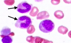
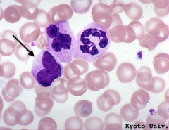
What kind of cells are this? How can you tell?
|

Band / Stab Cells
- Stage: 5 - Cell size: approximately same as mature cell - Nucleus: horseshoe-shaped / deeply indented (>1/2 of diameter) - Cytoplasm: pink granules |
|

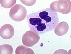
What kind of cell is this? How can you tell?
|
Mature Segmented Neutrophil
- Stage: 6 - Cell size: mature size - Nucleus: multiple nuclear lobes (3-5) separated by thin nuclear filament, called a "segment" - Cytoplasm: pink granules |
|
|
What is the pattern of maturation of eosinophils and basophils?
|
Similar way as neutrophils (all granulocytes)
1. Myeloblast (same for all) 2. Promyelocyte 3. Myelocyte 4. Metamyelocyte 5. Stab Cell 6. Eosinophil or Basophil |
|
|
How common are eosinophils vs basophils in bone marrow? Blood?
|
- Eosinophils are more commonly encountered in bone marrow
- Basophils are rarely encountered in normal bone marrows - Both are more common in blood |
|
|
Monocytes:
- Size - Cytoplasm - Nucleus |

- Size: large (2-3x RBC)
- Cytoplasm: abundant grey/blue cytoplasm w/ variably present, delicate pink granules which may be difficult to see in BM - Nucleus: reniform / kidney-bean shaped or folded nucleus, no nucleoli |
|
|
Where are monocytes more easily identified?
|
Blood by their abundant cytoplasm
|
|
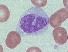
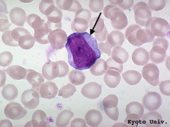
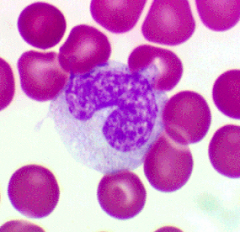
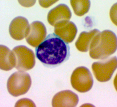
What kind of cell is this? How can you tell?
|

Monocyte:
- Size: large (2-3x RBC) - Cytoplasm: abundant grey/blue cytoplasm w/ variably present, delicate pink granules which may be difficult to see in BM - Nucleus: reniform / kidney-bean shaped or folded nucleus, no nucleoli |
|
|
Lymphocytes:
- Size - Cytoplasm - Nucleus |

- Size: smallest nucleated cell in BM, roughly same as RBC
- Cytoplasm: scanty pale blue cytoplasm - Nucleus: high nuclear to cytoplasmic ratio, uniformly condensed chromatin, no nucleoli |
|
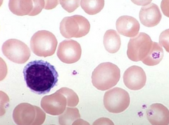
What kind of cell is this? How can you tell?
|

Lymphocyte
- Size: smallest nucleated cell in BM, roughly same as RBC - Cytoplasm: scanty pale blue cytoplasm - Nucleus: high nuclear to cytoplasmic ratio, uniformly condensed chromatin, no nucleoli |
|
|
Where do T cells mature? Where do they seed?
|
- Mature in thymus
- Seed blood and lymphoid organs |
|
|
Where do B cells mature? What are their progenitor cells called?
|
- Mature in bone marrow
- Progenitor cells are called Hematogones |
|
|
What are Hematogones?
|
B cell progenitor cells
|
|
|
What is the term for the formation of platelets?
|
Thrombopoiesis (platelets aka thrombocytes)
|
|
|
The CFU which produces platelets gives rise to what cells?
|
Megakaryocytes
|
|
|
Megakaryocytes:
- Size - Nucleus - Cytoplasm |

- Size: large (up to 100 µm diameter)
- Nucleus: single, multi-lobated - Cytoplasm: pink/gray, resembling platelets |
|

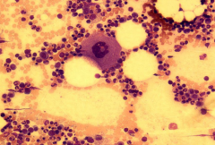
What kind of cell is this? How can you tell?
|

Megakaryocyte
- Size: large (up to 100 µm diameter) - Nucleus: single, multi-lobated - Cytoplasm: pink/gray, resembling platelets |
|
|
What is a routine site for bone marrow biopsies?
|
Iliac Crest
|
|

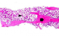
What can you see on a bone marrow core biopsy?
|

- Bone trabeculae (bright pink, thin, contain few osteocytes)
- Adipose tissue (large, clear vacuoles) - Hematopoietic elements (other purple cells) |
|

How do you determine the bone marrow cellularity?
|
Estimate the relative percentage of hematopoietic cells to fat
|
|
|
What does the bone marrow cellularity vary with? How can you estimate what it should be?
|
- Varies with age
- Very general rule: Normal Cellularity = 100 - Age |
|
|
What are the two parts of blood?
|

- Formed elements (including cells and platelets)
- Plasma |
|
|
What does plasma consist of?
|
- 90% water
- Electrolytes (Na+, Cl-, K+, PO4-, HCO3-) - Glucose - Amino Acids - Immunoglobulin - Complement Proteins - Steroids - Clotting Factors |
|
What is the term for the color intensity of normal RBCs? What determines the intensity?
|
Normochromatic (directly related to amount of hemoglobin)
|
|
|
What is the term for decreased color staining intensity of RBCs?
|
Hypochromasia
|
|
|
What is the term for increased color staining intensity of RBCs? What other feature?
|
Hyperchromasia (also have lack of central pallor)
|
|
|
In the correct viewing areas, most of the RBCs have a pale staining center. Why?
|

- RBCs have a biconcave shape
- Center of RBCs stains pale because there is less material in this thin part |
|
|
What is the function of the RBC's biconcave shape?
|
Maximizes the surface to volume ratio facilitating gas exchange
|
|
|
What cytoskeletal elements maintain the erythrocyte's shape?
|
Spectrin, Actin, Ankyrin
|
|
What is the term for RBCs with variation in cell size?
|

Anisocytosis
|
|

What is the term for RBCs with variation in cell shape?
|
Poikilocytosis
|
|

Based on the shape of this erythrocyte, can you identify this blood disorder? Cause?
|

Sickle Cell Disease - result of a hereditary hemoglobinopathy / structurally abnormal hemoglobin
|
|
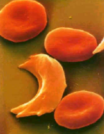
What symptoms would you expect to find in a patient with this abnormality?
|

Patients w/ severe Sickle Cell Disease have:
- Severe anemia (cells are fragile and lyse) - Vaso-occlusive complications (membranes adhere to endothelium causing narrowing of small vessels which traps sickled cells, leads to vascular occlusion, and ischemic tissue damage (painful crises) - Chronic hyperbilirubinemia (large amounts of bilirubin in blood, manifests as jaundice) |
|
|
Are nuclei visible in mature erythrocytes?
|
No
|
|
|
What effect does not having a nucleus have on a mature erythrocyte?
|
RBCs have a limited life span of approx. 120 days in circulation
|
|
|
What does it mean if a patient has increased reticulocytes in their peripheral blood?
|
Indicates an increased demand for erythrocytes
|
|
|
What conditions might produce the premature release of reticulocytes from BM into peripheral blood?
|
Reticulocytes may appear in blood as a result of:
- Hemorrhage → loss of RBCs - Anemia → loss of RBCs - Ascent to high altitude → increased O2 demand |
|
|
What is the size of dried RBCs?
|
7 µm
|
|
|
What are the types of granules in leukocytes? How does their presence determine the type of leukocyte?
|
- Non-specific (primary, azurophilic)
- Specific (secondary) - Granulocytes (neutrophils, eosinophils, and basophils) have non-specific AND specific granules - Monocytes and Lymphocytes lack specific granules but may contain non-specific granules |
|
|
Mature Neutrophils:
- Size - Proportion of WBCs - Nuclei |
- Size: 10 µm (slightly larger than RBCs)
- Proportion of WBCs: 30-70% - Nuclei: several lobes (3-5) connected by thin filaments |
|
|
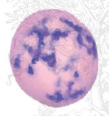
How do neutrophils differ if they are from females? Why?
|

- Females may have neutrophils with nuclear lobes that have a drumstick-shaped nuclear appendage
- Nuclear appendage is the inactivated X chromosome (Barr body) |
|

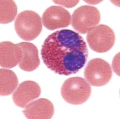
Who has neutrophils with this appearance? Why?
|
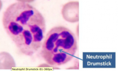
Females
- Nuclear appendage is the inactivated X chromosome (Barr body) |
|
|
What is the color of the neutrophil granules / cytoplasm?
|

- Neutrophilic granules do not stain w/ either basic or acidic dyes
- Similar to color of erythrocyte cytoplasm |
|
|
What kind of granules are in Neutrophils? How do they differ? Similar?
|

- Non-specific (primary) granules are fewer in number in normal granulocytes
- Specific (secondary) granules are more numerous - Both contain enzymes critical to neutrophil function |
|
|
What are the substances within non-specific granules in neutrophils?
|

Lysosomes containing:
- Acid hydrolases - Lysozyme - Other enzymes |
|
|
What are the substances within specific granules in neutrophils?
|

- Alkaline phosphatase
- Amino peptidase - Collagenase - Other enzymes |
|
What are the known functions of neutrophils?
|
- Phagocytosis
- Destruction of microorganisms - Initiation of inflammatory process |
|
|
Mature Eosinophils:
- Size - Proportion of WBCs - Nuclei - Cytoplasm |

- 10-14 µm (larger than neutrophils)
- 0-7% - Bi-lobed nucleus - Reddish granules w/ darker red or orange cytoplasm |
|
|
What do the specific (secondary) granules of Eosinophils contain?
|

- Major Basic Protein (MBP)
- Other basic proteins that combat parasites |
|
|
What are the functions of Eosinophils?
|
Increased in number in parasitic worm infections and allergic reactions
|
|
|
Why do Eosinophils stain red?
|

Large number of arginine residues in major basic protein give granules their eosinophilic staining property
|
|
|
Eosinophils function outside the circulation in what tissues? Why are eosinophils in the tissue spaces rather than in blood vessels?
|

- Can be found in the dermis of the skin and in CT components of the respiratory tree, GI tract, uterus, and vagina
- They are within tissue spaces so that they can encounter foreign microorganisms and antigens |
|
|
Mature Basophils:
- Size - Proportion of WBCs - Nuclei - Cytoplasm |
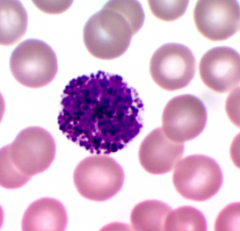
- 8-10 µm (small)
- <1% - Obscured irregular nucleus - Specific granules stain very dark purple and obscure nucleus |
|
|
How might antihistamines affect Basophils?
|
Anti-histamines inhibit degranulation of basophils
|
|
|
What kind of granules are found within monocytes and lymphocytes?
|
No specific (secondary) granules but may contain some primary granules
|
|
|
Monocytes:
- Size - Proportion of WBCs - Nuclei - Cytoplasm |
- 15-20 µm (largest WBCs)
- 5-12% - Large, eccentric, pale staining (bc of euchromatin) and indented to give horseshoe or S shape - Pale gray-blue cytoplasm and variably vaculated |
|
|
When monocytes enter peripheral tissues they differentiate into what?
|
Macrophages
|
|
|
What are the functions of Macrophages / Monocytes?
|
- Phagocytosis
- Antigen presentation on MHC molecules to T cells which are stimulated to respond |
|
|
Lymphocytes:
- Size - Proportion of WBCs - Nuclei - Cytoplasm |
- 5-15 µm
- 20-50% - Large, round, dark-staining nucleus that takes up most of volume - Cytoplasm is thin, blue rim |
|
|
The larger lymphocytes may be activated B cells which secrete what?
|
Antibodies
|
|
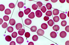
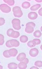
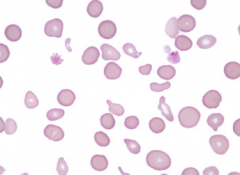
What are these structures? How can you tell?
|

Platelets
- Purple blue particles - Looks like debris - Smaller than RBCs, less numerous, and variably shaped |
|
|
Platelets are derived from large cells in BM called what?
|
Megakaryocytes
|
|
|
What is the function of platelets?
|
Involved in blood clot formation
|
|
|
How many nuclei are present in platelets? How many nuclei are present in megakaryocytes?
|
- Platelets: none
- Megakaryocytes: one |
|
|
What is Thrombocytopenia? Principal symptom?
|

- Condition in which there is an abnormally low number of platelets in peripheral blood
- Mucocutaneous bleeding |