Reading...
![]()
Play button
![]()
Play button
![]()
Use LEFT and RIGHT arrow keys to navigate between flashcards;
Use UP and DOWN arrow keys to flip the card;
H to show hint;
A reads text to speech;
50 Cards in this Set
- Front
- Back
|
|
|
|
|
|
|
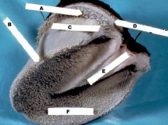
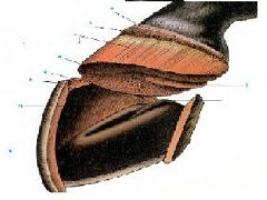
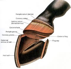
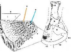
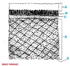
1. Stratum Externum
2. Stratum Medium 3. Stratum Internum 4. Hoof Wall 5. Dermis 6. Fused to periosteum of distal phalanx 7. PIII 8. PIV 9. PII 10. PI 11. Toe 12. Quarter 13. Heel |
|
|
|
|

What System/Organ is represented?
|
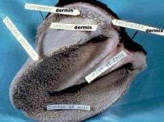
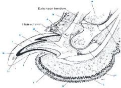
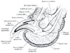
Integumentary/Claw
1. Claw Fold 2. Dermis 3. Haired Side 4. Distal Phalanx 5. Non-Keratinized Stratified Squamous Epithelium 6. Wall (Keratin) 7. Sole 9. Middle Phalanx |
|
|
|
|
|
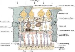
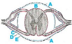
SENSITIVE RETINA 6/10 LAYERS
A. Axons of Ganglion Cells B. Ganglion Cell Layer C. Bipolar Cell Layer D. Cell Bodies of Rods&Cones E.Pigmented Epithelium F.Rod G. Cone H. Rods&Cones Layer |
|
|
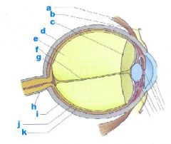
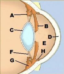
A-Canal of Schlemm
B-Iris C-Cornea D-Anterior Chamber E-Posterior Chamber F-Lens G-Ciliary Body H-Zonula I-Ora Serrata Covering Vitreous Space |
|
|
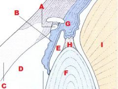
A-Ciliary Processes
B-Iris C-Lens D-Cornea E-Anterior Chamber F-Zonules G-Ciliary Body H-Trabecular Meshwork |
|
|
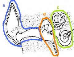
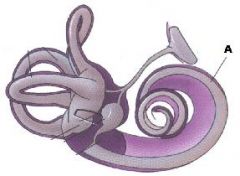
A-External Ear
B-Middle Ear C-Inner Ear |
|
A)Identify
B)What type of tissue? C)Function |
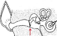
A. Tympanic Membrane
B. 3 Layers: -Dense CCT in middle -Lined on each side by epithelium C. Separates external ear from middle ear |
|
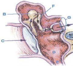
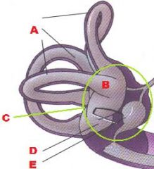
A) Identify Organ/Division and what are the bones called?
|
A)Middle Ear, 3 Auditory Ausicles
B)Malleus C)Tympanic Membrane D)Tympanic Cavity E)Auditory Tube F)Incus G)Vestibular Window H)Stapes |
|
|
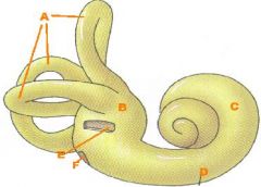
INTERNAL EAR
A. Semicircular Canals B. Vestibule C. Osseous Labrinth D. Cochlea E. Vestibular Window F. Cochlear Window |
|
|
INTERNAL EAR
A. Membranous Labrinth (Within Osseous Labrinth) |
|
|
INTERNAL EAR
A)Semicircular Canals B)Ampulla C)Vestibule D)Utricle E)Saccule |
|
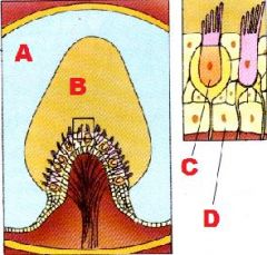
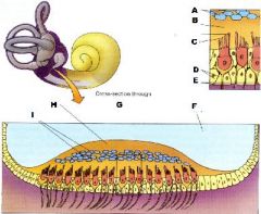
What is this a Picture of?
|
-Cross-section thru an Ampulla showing Crista Ampularis
A. Endolymph (in semicircular canal) B. Cupula C. Hair Cell with stereocilia & 1 Kinocilium D. Supporting Cell |
|
|

|
|
What is this?
|
-Embryo
A. Prosencephalon B. Mesencephalon C. Rhombencephalon D. Notochord |
|
|
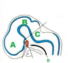
A. Cerebrum
B. Thalamus C. Midbrain D. Cerebellum E. Pons F. Medulla G. Spinal Cord |
|
|
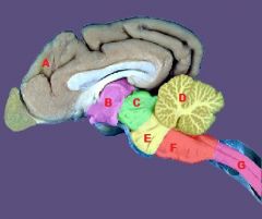
A. Prosencephalon = Cerebrum+Thalamus+Hypothal
B. Mesencephalon=Midbrain C. Rhombencephalon = Pons + Cerebellum + Medulla Oblongata |
|
|
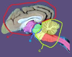
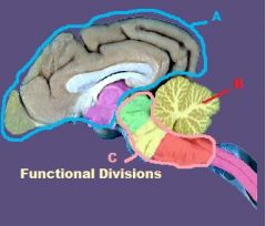
A. Forebrain
B. Cerebellum C. Brain Stem |
|
What organ?
|
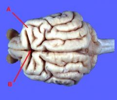
-Cerebrum
A.Gyri (Ridges-Gyrus singular) B.Sulci (Valleys-Sulcus) |
|
|
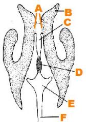
A. Central Canal (Lined with
ciliated columnar epithelium) B. Gray Matter C. White Matter D. White Matter E. Gray Matter |
|
|
|
|
|
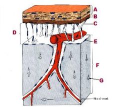
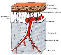
A. Subdural Cavity
B. Subarachnoid Cavity C. Pia Mater D. Arachnoid E. Dura Mater |
|
|
|
|
|
|
|
|
|
|
|
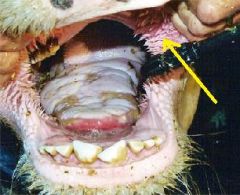
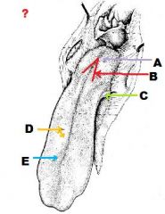
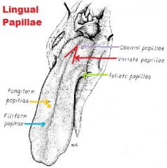
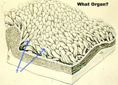
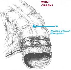
Conical Buccal Papillae
(Heavily Keratinized mucosal projections) (Ruminants) |
|
|
|
|
|
|
|
|
|
|
|
|
|
|
|
|
|
|
|
|
|
|
|
|
|
|
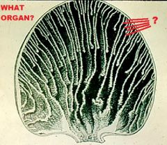
Interior of Rumen
Papillae |
|
|
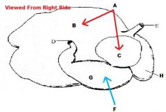
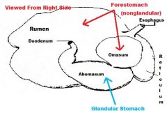
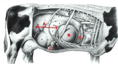
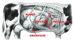
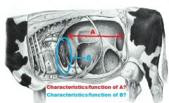
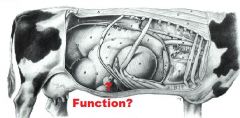
A) Rumen-opened up
- largest compartment - nearly fills left side of abdomen - large space for fermentation (chemical breakdown) B) Reticulum: -contats diaphr. Cr to rumen -mechanical breakdown of food |
|
|
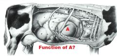
A. OMASUM
- located low in the cranial right abdomen - function is for mechanical breakdown of food |
|
|
Interior of Omasum
Omasal Laminae of different lengths |
|
|
Abomasum
- located ventrally & towards the right - glandular or “true” stomach; provides chemical digestion - has cardiac, proper gastric & pyloric gland regions - spiral folds in body regions; see grossly; are permanent |
|
|
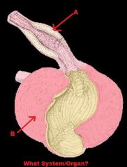
Avian Digestive System/Stomach
A- Proventriculus = glandular part B - Ventriculus (Gizzard) = Muscular Part |
|
|
|
|
|
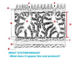
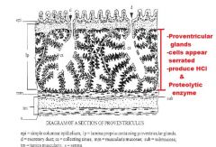
Ventriculus(Gizzard)
A. Koilin or Keratinoid B. Tubular Glands in LP C. Submucosa D. Tunica Muscularis (CCT) E. Serosa NO MUSCULARIS MUCOSAE! |
|
|
Large Intestine
A. Taenia coli - see in some species: Horses, pigs, humans -longitudinal bands of smooth muscle - very prominent in the horse |
|
|
|
|
|
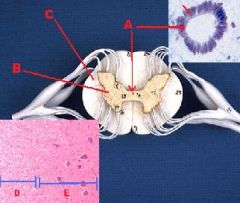
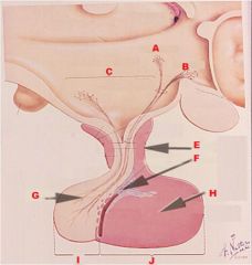
A. Paraventricular Nucleus
B. Supra-Chiasmatic Nucleus C. Hypothalamo-Hypophyseal Portal Tract E. PT F. PI G. PN H. PD I. Neurohypophysis J. Adenohypophysis |
|
|
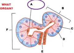
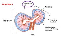
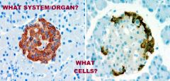
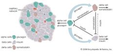
Endocrine System
Pancreas Alpha Cells -Secrete Glucagon |
|
|
Nice
|