![]()
![]()
![]()
Use LEFT and RIGHT arrow keys to navigate between flashcards;
Use UP and DOWN arrow keys to flip the card;
H to show hint;
A reads text to speech;
28 Cards in this Set
- Front
- Back
|
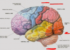
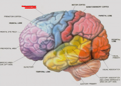
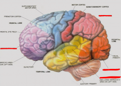
Association areas of cerebral cortex |
Responsible for recognition, comprehension, cognition, i.e. executive function, memory, and language Integrate multiple sensory and motor modalities Located adjacent to primary and secondary sensory and motor areas |
|
|
Sensory cortices - primary auditory, visual, somatosensory Output - motor cortex - Upper motor neurons that drive LMNs and muscle |
|
|
Supplementary motor area in premotor cortex Areas directly associated with motor cortex - cusp between primary and association cortex. |
|
|
Prefrontal area - complex cognitive function Visual association area Auditory association area Parietal lobe - integrates somatosensory and visual information |
|
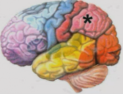
Parietal association area |
Adjacent to primary somatosensory, auditory, and visual cortices. Involved with spatial sensation and manipulation of objects in space Contralateral - right parietal controls left side and vice versa Duties - Stereognosis, body image |
|
|
Stereognosis |
Ability to recognize objects by touch alone. Lesions = astereognosis Localized in parietal association area |
|
|
Body image |
Conciousness of body's position at any time. Lesions cause neglect syndrome - iIgnoring of contralateral side of body and associated surrounding space. Do not draw contralateral visual field, eat food there, etc. |
|
|
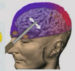
Rubber hand illusion |
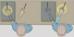
Hide real hand, show rubber hand so sensory information from real and visual from rubber. Patient freaks out if threaten to smash rubber hand with hammer. Visual system overrides somatosensory input. Area in prefrontal, not parietal cortex, is responsible for this illusion. |
|
|
Prefrontal association areas |
Anterior to and projects to premotor/motor areas Lesions in monkey results in delayed response (spatial working memory) |
|
|
Delayed alternation test |
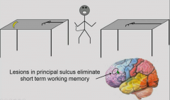
Experimental paradigm for prefrontal association areas. Monkey sees food that is out of reach. Give monkey short rake, long rake, and banana. Monkey without lesion uses short rake to retrieve long rake to retrieve banana. Lesion in principal sulcus eliminates ability to perform task by eliminating short term working memory - forgets about long rake when looking at short rake. |
|
|
Non-alternating test |
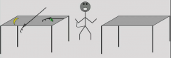
Short rake, long rake, and banana on same table. No short term memory required. Principal sulcus lesioned and non lesioned monkey perform task the same. |
|
|
Where does short term working memory reside? |
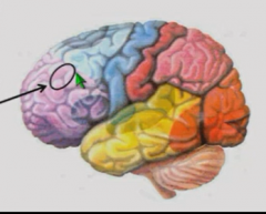
Principal sulcus "Brain's chalkboard" Neurons here map out contralateral visual space, and continue to fire after visual cue is gone for short period of time. |
|
|
Limbic association areas |
Cingulate gyrus, orbitofrontal cortex, anterior temporal cortex (Hippocampus) Connected to hippocampus, amygdala that have strong emotional content. |
|
|
Lesions of anterior temporal cortex |
This is limbic cortex and includes hippocampus, leads to profound deficients in long-term memory. |
|
|
Lesions of orbitofrontal cortex |
Calming effect, decrease aggressiveness. Lobectomies of this area in schizophrenia patients resulted in patients becoming docile and institutionalized. Frontal lobotomy - cut connections between orbitofrontal cortex and rest of brain. Go through eye socket. Ended with discovery of antipsychotic drugs in 1960s. |
|
|
Emotional memory |
Stored in limbic association areas (orbitofrontal cortex, hypothalamus) and sensed by amygdalas If emotional investment in event, remember better - e.g. food, hot burner. |
|
|
Orbitofrontal cortex and marketing |
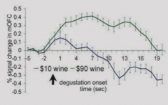
Patient connected to fMRI and fed wine in tube after being told whether wine is cheap or expensive. If told more expensive wine, like it more. Orbitofrontal cortex is active for longer period of time. If told it is cheap wine, like it less, and orbitofrontal cortex declines in activity. If don't tell subject price, can't tell the difference. |
|
|
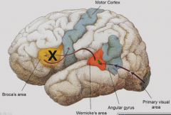
Wenicke-Geschwind model |
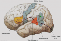
Language is localized in 95% of people in left side of brain. In reading, primary visual information goes through primary visual area to angular gyrus where words are encoded from lines. (If repeating, begins with primary auditory area). Then, to Wernicke's area, where auditory and visual information is integrated and comprehension takes place. If reading aloud, Wernicke's area projects to Broca's area (via arcuate fasciculus) which controls UMNs in motor cortex to control speech muscles. |
|
|
Broca's area |
Frontal lobe premotor area. Controls speech muscles and contains programs for fluent speech |
|
|
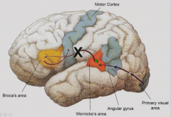
Arcuate fasciculus |
Pathway connecting Broca's and Wernicke's area |
|
|
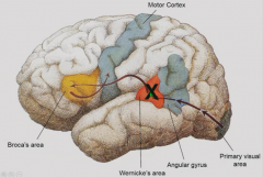
Wernicke's area |
Superior temporal/inferior parietal junction. Language comprehension and intent to initiate spoken language. |
|
|
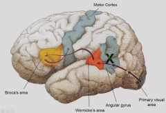
Angular gyrus |
Integrates visual input for reading to language centers. |
|
|
PET - Areas active when just listening, speaking, generating verbs, and seeing words |
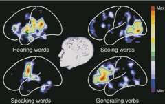
Listening - Wernicke's area and auditory cortex Speaking - add Broca's and motor cortex Generating verbs (creativity) - more activity in Broca's Seeing words - additional activity in visual cortex |
|
|
Aphasia |
Defects in comprehension of production of language due to injury or stroke on left side of brain. |
|
|
Broca's aphasia |
Damage to Broca's area Expressive aphasia - slow, halting speech Comprehension is intact. |
|
|
Wernicke's aphasia |
Damage to Wernicke's area Cannot comprehend written or heard language or communicate to Broca's area what it intends to say. Broca's problems are out of control - spoken language that is seemingly fluent but without content - "word salad" |
|
|
Disconnection/conduction aphasia |
Damage to arcuate fasciculus Able to comprehend spoken or written language (Wernicke's area is intact) Because Wernicke's cannot control Broca's, speaks in word salad. |
|
|
Alexia |
Lesion in angular gyrus Affects reading while sparing other components of language. |