![]()
![]()
![]()
Use LEFT and RIGHT arrow keys to navigate between flashcards;
Use UP and DOWN arrow keys to flip the card;
H to show hint;
A reads text to speech;
143 Cards in this Set
- Front
- Back
|
Neuroglia |
Glial cells. Supports and protect neurons. These outnumber neurons 10:1. No nerve transmission. |
|
|
Myelinated axons |
Conduct impulses faster than un_______ axons. Form by different glial cells depending on location. Ogliodendrocytes are in the brain and spinal cord, and Schwann cells are in the peripheral nerves. |
|
|
Nodes of Ranvier |

Gaps in the myelin sheath that allow faster nerve impulse conduction due to 'skipping'.( Saltatory conduction). |
|
|
Saltatory Conduction |
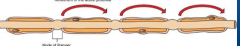
Rapid means of conduction an action potential along myelinated axons. Depolarization only happens at the Nodes of Ravier.Allow vision and fine motor skills. Multiple sclerosis |
|
|
Multiple sclerosis |
Damages to myelin sheath that slows the muscle response. Muscles don't respond to what brain wants to do. |
|
|
Organization of the Nervous System |
Anatomical, Direction of impulses, and function. |
|
|
Central Nervous System |
Anatomical nervous system. Brain and spinal chord. Along the central axis of the body. |
|
|
Peripheral nervous system |
Anatomical Nervous System.Extends outward from the central axis toward the periphery of the body. Cranial nerves originate directly from the brain. Spinal nerves emerge from the spinal chord. |
|
|
Afferent Nerves |
Direction of impulses. Take impulses to the CNS. Sensory nerves . |
|
|
Efferent Nerves |
Direction of impulses. Takes impulses away from the CNS. Motor nerves to target organs. May carry fibers that are sensory, motor, or both signals. |
|
|
Somatic Nervous System |
Function. Actions under conscious, or voluntary control. Impulses sent mainly to skeletal muscle. |
|
|
Autonomic Nervous System |
Controls and coordinates involuntary functions. Ex. Slowing of the heart rate in response to an increased blood pressure. Impulses send to smooth and cardiac muscles and glands. |
|
|
Neuron Function |
Resting membrane potential. High Na outside, low inside. High K+ inside, low outside. |
|
|
Action Potential |
Nerve impulse. Depolarization and repolarization |
|
|
Depolarization |
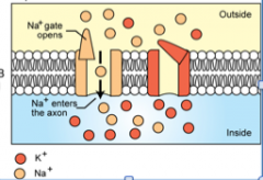
When the neuron receives a stimulus. Na+ channels open in the membrane. Na+ flows into cell by passive diffusion. The inside of the cell becomes +. |
|
|
Repolarization |
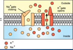
Na+ channels close, K+ channels open as the K+ flows out of the cells. Inside of the cell is negative again, bringing it back to resting potential. |
|
|
Generation of an Action Potential |
Na+/K+ pump restores the ions on the correct side of the membrane. Outside is + and inside is -. Maintained by the pump by pumping Na+ from the inside of a neuron to the outside, and K+ from the outside to the inside. Active transport. |
|
|
Threshold Stimulus |
Stimulus that is strong enough to make the neuron response and cause completely depolarization. The all or nothing principle. |
|
|
Once an action potential in generated |
Depolarization spreads down membrane in a wave. "Nerve impulse". |
|
|
Communal nerve |
A bundle of axons |
|
|
Resting membrane potential |
Difference in electrical charge across the membrane. From differences in distribution of positive and negative ions including potassium, proteins, and other charged ions. |
|
|
Muscle cells |
modified to respond to electrical signals. |
|
|
electricity |
All communication from your body comes from ___________. |
|
|
Nervous system |
Look at slide 8 |
|
|
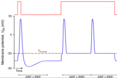
Action potential graph |

|
|
|
Threshold Stimulus and sustained threshold stimulus |
|
|
|
Synaptic Transmission |
Communication between neurons. Involves the synapse, the synaptic cleft, presynaptic neuron and postsynaptic neuron. |
|
|
Synapse |
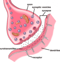
Junction between two neurons, or a neuron and a target cell. |
|
|
Synapse |

|
|
|
Synaptic cleft |
Gap between adjacent neurons |
|
|
Presynaptic neuron |
Brings the depolarization wave to the synapse, and releases the neurotransmitter |
|
|
Postsynaptic neuron |
Contains receptors for the neurotransmitter. |
|
|
Step 1 in the Neurotransmitter Action |

Neurotransmitter molecules are synthesized from precursors under the influence of enzymes . |
|
|
Step 2 in the Neurotransmitter Action |

Neurotransmitter molecules are stored in vesicles |
|
|
Step 3 in the Neurotransmitter Action |
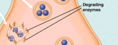
Neurotransmitter molecules that leak from vesicles are destroyed by enzymes |
|
|
Step 4 in the Neurotransmitter Action |
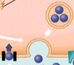
Action potential causes vesicles to fuse with the presynaptic membrane and release their neurotransmitter molecules into the synapse. |
|
|
Step 5 in the Neurotransmitter Action |
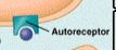
Released neurotransmitter molecules bind with autoreceptors and inhibit subsequent neurotransmitter release . |
|
|
Step 6 in the Neurotransmitter Action |

Released neurotransmitter molecule bind to postsynaptic receptors. |
|
|
Step 7 in the Neurotransmitter Action |

Released neurotransmitter molecules are deactivated either by reuptake or enzymatic degradation. |
|
|
Acetylcholinesterase |
Mono-amine oxidase (MAO). Antidepressants prevent breakdown and reabsorption of neurons. Degrades the synaptic transmitters. Either excitatory or inhibitory on target cell, depending on the location. Main transmitter for the parasympathetic nervous system. Neurotransmitter. |
|
|
Examples of acetylcholinesterase |
Excite motor neurons, inhibits heart rate. |
|
|
Catecholamines |
Excitatory neurotransmitter. Norepinephrine and Epinephrine (basically the same). Produced in top concave portion of the kidneys. Made in the adrenal gland in the sympathetic nervous system. |
|
|
Dopamine |
Type of neurotransmitter that is involved with automatic functions and muscle control. Found in the brain. Associate with tremors in Parkinson's disease . |
|
|
Parkinson's Disease |
Causes tremors. Lack of dopamine neuron function. |
|
|
Gamma-aminobutyric acid (GABA) |
Type of inhibitory neurotransmitter in the brain. |
|
|
Glycine |
Type of inhibitory neurotransmitter in the spinal cord |
|
|
Valium |
Type of glycine tranquilizer that works by increasing GABA effects on brain. Tranquilization leads to sedation. |
|
|
Central Nervous System |
Consist of the spinal and cord and brain, which includes the cerebrum, cerebellum, diencephalon, and the brain stem. |
|
|
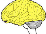
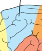
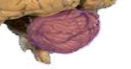
Cerebrum |
Part of the brain responsible for higher order behaviors including learning, intelligence, and awareness. Consists of gyri and sulci, longitudinal fissure, gray matter, and white matter. Telecephalon. |
|
|
Cerebrum |
Folded to allow more surface area to allow more neurons, to allow more synapses, to allow more neural ability |
|
|
Frontal lobe |

Lobe of the cerebrum where voluntary movement planned, psychomotor skills, motor cortex. |
|
|
Parietal lobe |
Location of pain, touch, temperature, and perception in the cerebrum. Mostly sensory information. |
|
|
Parietal ; frontal |
________ lobe senses pain in the hand, then sends signals to the _______ lobe that figures out what needs to be done to remove pain from the hand. |
|
|
Occipital lobe |
Visual interpretation in the cerebrum |
|
|
Temporal lobe |
Auditory, behavior, and memory in the cerebrum |
|
|
Cerebellum |
Located caudal to the cerebrum. The area of the brain responsible for coordinated movement, balance, posture, and complex reflexes |
|
|
Hypoplastic Cerebellum |
Motor coordination that is not formed correctly. Subject wobbles when they move. |
|
|
Diencephalon |

Passageway between the brain and the cerebrum. Includes the hypothalamus, thalamus, and the pituitary |
|
|
Thalamus |

Regulates sensory inputs to the cerebrum (bouncer) |
|
|
Hypothalamus |

Interface between the nervous system and the endocrine system. |
|
|
Pituitary |
Endocrine, the master gland. Rests in the pituitary fossa. Produces hormones that produces the body's ciruclation. Growth hormones, water intake, etc. |
|
|
Brain stem |
Connection between the rest of the brain and the spinal cord. The most primitive part of the brain. Include the medulla oblongata (caudal part), pons, and midbrain. Basic support functions of the body including heart beat, respiration, etc. Many cranial nerve originate here. |
|
|
Brain stem |

|
|
|
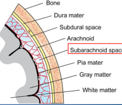
Meninges |
Connective tissue layer that surrounds the brain and spinal cord. Composed of three layers: dura mater, arachnoid, and pia mater. |
|
|
Gray matter |
Part of the cerebrum located in the cerebral cortex, which is the outer layer of the brain that consists of neuron cell bodies. |
|
|
White matter |
Communication in the cerebrum, how all the signals travel from the neurons to all the places they have to go. Have myelinated axons and the corpus callosum |
|
|
Myelinated axons |
Fibers beneath the cortex in white matter. |
|
|
Corpus callosum |
Fibers that connect the two halves of the cerebral cortex and allow communication in white matter. |
|
|
Midbrain |
Controls basic autonomic functions in the brain stem. |
|
|
Outline of skull layers |
|
|
|
Cerebrospinal Fluid |

Circulates around the brain (subarachnoid space), within ventricle system, and within the spinal canal. Clear, colorless, and has low protein. Made by the choroid plexus in 4th ventricle. |
|
|
Functions of the cerebrospinal fluid |
Protection and buoyancy, circulate nutrients, and removes waste products. |
|
|
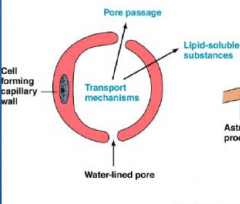
Blood Brain Barrier |
Separates body's blood supply in the brain from the nervous tissue. Capillary walls that have no fenestrations. Prevent many drugs, proteins, ions, and other molecules from readily passing from the blood into the brain. |
|
|
Capillary walls in the brain |
Consists of endothelial cells connected by tight junctions. Covered process of glial cells. |
|
|
Most capillaries in the body |
|
|
|
Brain capillaries |

|
|
|
12 |
___ Nerve pairs in the brain originate directly from the brain. Each may carry sensory signals, motor signals, or both. |
|
|
Cortex |
The outer part of the spinal cord that consists of white matter (axons). |
|
|
Medulla |
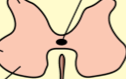
Inner part of the spinal cord (gray matter). Includes the central canal which contains CSF. Middle portion where the neurons are. |
|
|
Dorsal nerve roots |

Contain incoming sensory (afferent) fibers. Emerges between each pair of adjacent vertebrae. |
|
|
Ventral nerve roots |

Contain motor (efferent) fibers. Emerges between each pair of adjacent vertebrae. |
|
|
Ganglion |
Collection of neurons . |
|
|
Somatic reflexes |
Reflex that involves the contraction of skeletal muscles |
|
|
Autonomic reflexes |
Reflexes that regulate smooth muscle, cardiac muscle, and endocrine glands. |
|
|
Contralateral reflex |
Reflex that starts on one side of the body and travels to the opposite side. |
|
|
Ipsilateral reflex |
Reflex where the stimulus and response are on the same side of the nody. |
|
|
Relfex arc |
A basic process that all reflexes have. see slide 29. |
|
|
Stretch reflex |
includes a monosynaptic reflex arc, and sensory receptor is a muscle spindle, which when stretched, sends a signal causing the muscle to contract. Example is the patellar reflex. Antagonist muscle gets signaled to relax. Slide 31. |
|
|
Monosynaptic reflex arc |
Part of the stretch reflex. Involves a sensory neuron and a motor neuron. No interneurons involved. |
|
|
Crossed Extensor Reflex |
Contralateral reflex. Afferent sensory neuron synapses with interneurons that transmit information to the opposite side of the spinal cord. Causes contraction of opposite extensor muscles. Both sides of the body. |
|
|
Crossed extensor reflex |
When you step on a nail and raise your foot, the unaffected leg bears the weight. |
|
|
Hearing |
Converts vibration of air molecules into nerve impulses are that interpreted by the brain as sound. |
|
|
temporal |
Ear structures are mostly located in the ________ bone. |
|
|
External ear |
Part of the ear that is a funnel to collect sound wave vibrations and direct them into the rest of the ear. Have the pinna and auditory canal. Made of elastic cartilage. |
|
|
Middle ear |
Part of the ear that amplifies and transmits the vibrations to the inner ear. Has the tympanic membrane and auditory ossicles. |
|
|
Inner ear |
Contains the sensory receptors that convert the mechanical vibrations to nerve impulses. Also has receptors for equilibrium part of the inner ear. |
|
|
Cochlea |

Sensory receptor in the inner ear that converts the mechanical vibrations to nerve impulses. Cochlear duct. Involves the endolymph and sensory hair cells including "Organ of Corti" and Cochlear Nerve. Surrounded by Perilymph (U-shaped tube) |
|
|
Sensory hair cell |
Sit on the basilar membrane and tops the tectorial membrane in the cochlea. |
|
|
Step 1 in sound transmission |
Sound waves are collected by the pinna, and are funneled into the auditory canal. |
|
|
Step 2 in sound transmission |
Sound waves vibrate the tympanic membrane, which vibrates the three auditory ossicles attached to membrane. Ossicles vibrate the oval window at the entrance of the cochlea. |
|
|
Step 3 in sound transmission |
Vibration of the oval window vibrates the fluid in the cochlea. Sensory hairs moved up and down by fluid wave, and tips of hair hit tectorial membrane and get bent. |
|
|
Step 4 in sound transmission |
Bending of the hairs causes cochlear nerve to send a nerve impulse (auditory signal) to the brain. |
|
|
Equilibrium |
Maintains balance by keeping track of the position and movements of the head. Involves ________ receptors, information from the eyes, and proprioceptors throughout the body. has receptors in the vestibule and semicircular canals. |
|
|
Vestibule |
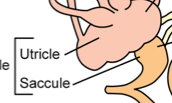
Part of the inner ear involved in equilibrium that has receptors with linear motion and static head position. Have the untricle and saccule. Has gelatin with otoliths on top of sensory hairs. |
|
|
Untricle and saccule |
Patches of sensory hair in each area of the vestibule. |
|
|
Semicircular canals |

Part of the inner ear involved in equilibrium that has receptors. Has rotational movement. Has 3 fluid tubs arranged in the X, Y, and Z axis. Has the ampulla, but doesn't have otoliths. |
|
|
Ampulla |
Sensory hair structure in each area of the semicircular canals. |
|
|
Nerve supply |
Vestibulocochlear nerve. |
|
|
Eye |
Various structures to control and focus light onto sensory photoreceptors. They convert electromagnetic energy into nerve impulses. Has the optic nerve. |
|
|
externally ; internally |
Both eyelids lined by haired skin ___________ and mucous membrane ____________
|
|
|
Superior palpebra |
More mobile of the eyelids and has cilia. |
|
|
Palabrae |
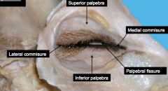
Superior palpebra, medial commisure, palpebral fissure, inferior palpebra, lateral commisure. |
|
|
Lacrimal glands |
Makes and secretes tears, and washes the surface of the eye. Drains into the nasolacrimal duct (nasal passage). |
|
|
Nictating membrane |
Cartilage plate. Lies medial between palpebrae and globe (eyeball). Has lymph modules and accessory lacrimal gland. |
|
|
External fibrous coat |
Layer of the globe (eyeball) including the cornea and sclera. Most superficial layer that completely surrounds the globe. Dense c.t. |
|
|
Middle vascular coat |
Uvea, layer of the globe (eyeball) including the iris, choroid, and ciliary body. Provides nourishment to the globe, controls pupillary diameter, and controls the shape of the lens. |
|
|
Internal coat |
Nervous tunic. Layer of the globe (eyeball) that includes the retina, and the nerves and vessels associated with it. |
|
|
Limbus |
Corneroscleral junction in the external fibrous coat. |
|
|
Cornea |
Takes up 1/4 of the anterior portion of the external fibrous coat. Transparent and avascular. |
|
|
Sclera |
Takes up 3/4 of the posterior portion of the external fibrous coat. Opaque and has elastic fibers. |
|
|
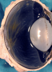
Iris |
Have two layers of smooth muscle, the circular (constrict) and radial (dilate), in the middle vascular coat of the eye. Located between the cornea and lens, controls pupil size, and pupil shape varies with species. |
|
|
Types of pupils |

|
|
|
Granulae iridicae of equine pupillary margin |
|
|
|
Horses and goats |
Horizontal shaped pupils |
|
|
Ciliary body |

Part of the middle vascular coat with muscle bundles covered by epithelium (ciliary processes). Ciliary muscles control lens shape: contract (rounder) and relax (flattens) |
|
|
Parts of middle vascular coat |

|
|
|
Choroid |
Middle vascular coat that contains melanin and the tapetum lucidum |
|
|
Choroid |

|
|
|
Tapetum lucidum |
Superior to the optic disc. Not in humans or pigs. |
|
|
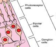
Retina |
Part of the internal coat. Contains the photoreceptors and the optic disc. Multiple layers of neutral cells (photoreceptor cells). |
|
|
Optic disc |
Blind spots; where axons from the retina leave the eye. Retinal vessels enter and leave here. |
|
|
Photoreceptors |
Bipolar cells and ganglion cells process the information from the _______________ . |
|
|
Rods |
Photorecepting cell that are low, light gray. |
|
|
Cones |

Bright-light, color. No fovea centralis in domestic animals. |
|
|
Sensory retina |
|
|
|
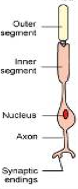
Neurons |
Basic function unit of the nervous system with a high requirement for oxygen. cannot reproduce but generate the cell process if the body remains intact. Have neuroglia. Has a cell body (soma or perikaryon) and dendrites (receive stimuli). Have axons and a high metabolic rate. |
|
|
Axon |
Conducts nerve impulses away. Single long process that may extend several feet. Some are myelinated. Have nodes of Ranvier. |
|
|
Anterior chamber |

Anterior portion of the chambers of the globe. Cornea to iris. 1. |
|
|
Posterior chamber |
Anterior portion of the chambers of the globe. Iris to lens. 2. |
|
|
Posterior position |
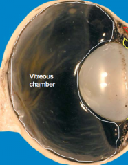
Vitreous chamber (body). |
|
|
Cornea, aqueous humor, lens, virtuous humor |
Light strikes the eye and gets refracted by ______, ___________________, ____ , _________________. |
|
|
Cornea; lens |
The ______ does most of the refraction, while the ______ only fine tunes the focus. |
|
|
When light converges onto the retina |
Photoreceptors stimulated. Nerve signal sent to brain via optic nerve. "Image" is actually sent upside down. Occipital lobe does initial processing. |