Reading...
![]()
Play button
![]()
Play button
![]()
Use LEFT and RIGHT arrow keys to navigate between flashcards;
Use UP and DOWN arrow keys to flip the card;
H to show hint;
A reads text to speech;
19 Cards in this Set
- Front
- Back
|
Describing Inner and outer segments photoreceptors
• orientation/direction of segments • the segment that contains the visual pigment • the segment that forms synapses and which cells it synapses with (2) • which segment experiences turnover |
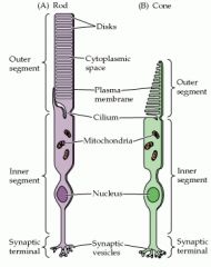
Describing Inner and outer segments photoreceptors:
• outer segment is directed towards the Retinal Pigment Epithelium (RPE); Inner segment is directed towards the eyeball • The outer segment contains the visual pigment • The Inner segment forms synapses w/ bipolar and horizontal cells • New disks are added to the base of the outer segment while older disks are displaced upward, shedded at the tip and phagocytosed by RPE cells |
|
|
Properties of Rods and Cones:
• Which are high/least sensitive to light • Which are best fxning under low/high light condition • Which has better temporal resolution • The number of types of cones • Which has lower spatial resolution and what causes this attribute • The names of the synapses of their inner segments • The neurotransmitter that is released and the light condition it is released in |
Properties of Rods and Cones:
• Rods are more sensitive to light than cones • Rods fxn better under low light conditions • Cones have better temporal resolution • There are types of cones • Rods have lower spatial resolution due to the basic wiring mechanism called convergence • Rod spherules, Cone pedicles • Glutamate, released in the dark |
|
|
The Visual pigment of rods:
• Name • Its two components - their origins |
The Visual pigment of rods:
• Rhodopsin • Two components - - Opsin, synthesized in the photoreceptor - Retinal, a chromophore derived from vitamin A and found in BOTH rods and cones |
|
|
Opsin:
- Location of molecule w/in the photoreceptor - General structure - relationship w/ Retinal |
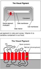
Opsin:
- Located in the disk membrane of photoreceptors - 7 transmembrane domains w/ amino terminal located in the disk interior and the carboxy group in the photoreceptor cytoplasm - Retinal is covalently attached to the 7th domain |
|
|
The Dark Current:
• State of photoreceptors in the dark • Name of the G-protein involved • Role of cGMP in the dark |
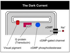
The Dark Current:
• They are depolarized • tranducin • It keeps photoreceptors depolarized through the opening of cGMP-activated Na+ channels |
|
|
The first step in the phototranduction process
|
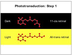
The visual pigment absorbs light and converts 11-cis retinal to all trans retinal, triggering the other steps of signal transduction
|
|
|
The 2nd step in Phototransduction:
Discuss the enzyme involved, what activates it, its substrate, its metabolite and the end result |
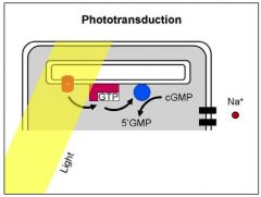
2nd step in phototransduction:
• The activated visual pigment with Transducin, activates PDE • PDE breaks down cGMP to 5' GMP • decreasing [cGMP] closes cGMP Na+ channels, hyperpolarizing photoreceptors |
|
|
Rods are insensitive to color but are they more sensitive to certain wavelengths of light or are they equally stimulated but all wavelengths?
|
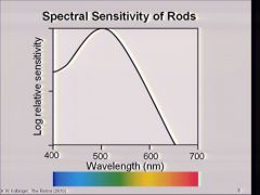
They have different sensitivities to wavelengths of light
|
|
|
The three types of retinal cones:
• Name - Associated color - maximum wavelength of sensitivity |
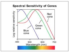
• S-cones
- "Blue" - 430nm • M-cones - "Green" - 530nm • L-cones - "Red" - 560nm |
|
|
Explain how all 3 cones types contribute to color vision
|
Perception of one color is not based on exclusive activation of a single cone but due to a pattern of stimulation and relative hyperpolarization of all 3 cones
|
|
|
Wiring of the retina:
• Two basic wiring systems - Name the types of retinal cells associated with each and onto which cells each synapse • Kinds of potentials elicited by retinal cells |
Wiring of the retina:
• Direct wiring system - photoreceptor cells synapse onto bipolar cell cells which synapse onto retinal ganglion cells associated with each and onto Indirect wiring system - The horizontal cells (outer plexiform layer) synapse onto bipolar cells, regulating input from photoreceptors - Amacrine cells each regulate the output of bipolar cells • All retinal cells synapsing in the retina produce graded potentials. Photoreceptors which send their long axons to the LGN, produce an AP |
|
|
Describe the similarities/differences btwn the types of bipolar cells, including:
- their response to light - the type of receptors on their surface - the type of synapse they have |
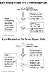
• ON-center bipolar cells:
- are depolarized by light - have ionotropic glutamate receptors - described as having a "sign-conserving" synapse since photoreceptors need to be hyperpolarized to excite them • OFF-center bipolar cells: - are hyperpolarized by light - have metabotropic glutamate receptors - described as having a "sign-converting" synapse since photoreceptors need to be hyperpolarized to inhibit them |
|

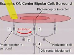
Explain the process of lateral inhibition with Photoreceptors, horizontal cells and ON-center bipolar cells when light is shone on the periphery of the ON Center bipolar cells
|

• When light is shone in the surround, the photoreceptor hyperpolarizes, reducing the release of the excitatory neurotransmitter, Glu
• Photoreceptors in the periphery of the visual field for ON-center bipolar cells synapse with inhibitory cells called Horizontal cells. This is a sign-conserving synapse. • The decrease Glu release causes Horizontal cells to hyperpolarize. Since hyperpolarization does not allow Horizontal cells to release their inhibitory NT, the cell they synapse with, the terminal end of photoreceptor cells, are then depolarized. This is a sign-converting synapse. • The depolarized photoreceptor cells then release Glu which inhibit the ON-center bipolar cell in a sign-converting synapse. |
|
|
Explain the process of lateral inhibition with Photoreceptors, horizontal cells and ON-center bipolar cells when light is shone on the periphery of the ON Center bipolar cells
|
|
|
Explain the process of lateral inhibition with Photoreceptors, horizontal cells and ON-center bipolar cells when light is shone on the periphery of the ON-center bipolar cells
*remember the types of synapses associated with each |
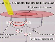
• When light is shone in the surround, the photoreceptor hyperpolarizes, reducing the release of the excitatory neurotransmitter, Glu
• Photoreceptors in the periphery of the visual field for ON-center bipolar cells synapse with inhibitory cells called Horizontal cells. This is a sign-conserving synapse. • The decrease Glu release causes Horizontal cells to hyperpolarize. Since hyperpolarization does not allow Horizontal cells to release their inhibitory NT, the cell they synapse with, the terminal end of photoreceptor cells, are then depolarized. This is a sign-converting synapse. • The depolarized photoreceptor cells then release Glu which inhibit the ON-center bipolar cell in a sign-converting synapse. |
|
|
ON-center ganglion cells:
• Have these kinds of synapses/connections with ON-center bipolar cells • generate these kinds of potentials • result when light is shone on the center of its receptive field |
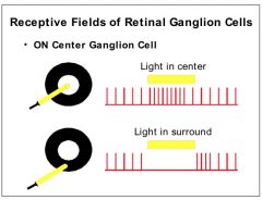
ON-center ganglion cells:
• sign-conserving/excitatory • APs • The frequency of APs increase |
|
|
OFF-center ganglion cells
• Have these kinds of synapses/connections with OFF-center bipolar cells The area of its receptive field that would need to be stimulated in order to increase the frequency of APs |
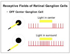
OFF-center ganglion cells
• Sign-conserving/excitatory • Stimulate the periphery |
|
|
Retinitis Pigmentosa:
• Mode of hereditary (3) • These cells usually degenerate • Earliest sx (2) • is associated with the lack of fxning of this process |
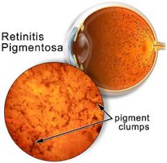
Retinitis Pigmentosa:
• Autosomal dominant & recessive, X-linked recessive • Rods usually degenerate • Night blindness followed by loss of peripheral vision, leading to tunnel vision • Reduced phagocytosis by RPE cells during the process of disk shedding |
|
|
Color Blindness:
• Most common form • Two types of the above • Name of test used to assess the condition |
Color Blindness:
• Red-Green • Two types of Red-Green colorblindness - Protanopia: L-cone is absent - Deuteranopia: M-cone is absent • Pseudoisochromatic color plate |