Reading...
![]()
Play button
![]()
Play button
![]()
Use LEFT and RIGHT arrow keys to navigate between flashcards;
Use UP and DOWN arrow keys to flip the card;
H to show hint;
A reads text to speech;
74 Cards in this Set
- Front
- Back
- 3rd side (hint)
|
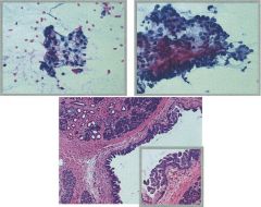
Amylase, lipase, leuk esterase in pseudocyst? Neoplastic?
Tumor markers in mucinous cyst? |
all high in psuedocyst.
all low in neoplastic cyst. Mucinous cyst has elevated ***CEA, CA19.9, CA125, CA15.3 IPMN can have elevated amylase. LOW LEVEL excludes pseudocyst |
|
|
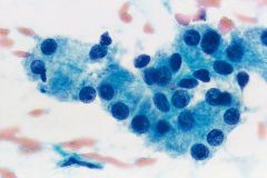
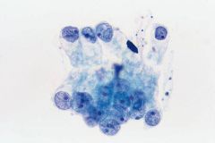
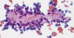
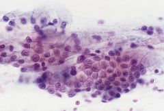
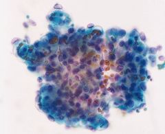
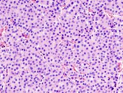
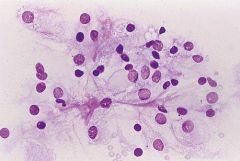
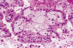
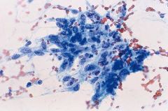
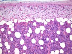
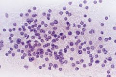
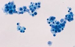
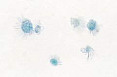
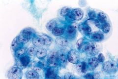
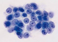
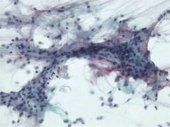
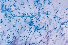
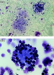
Pancreas FNA
|
Normal acini
Acinar arrangement Eccentric round nucleus Even chromatin, no necleolus Granular cytoplasm Indistinct cell borders |
|
|
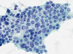
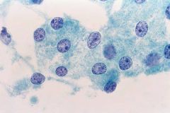
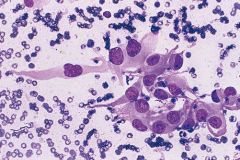
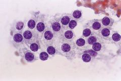
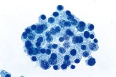
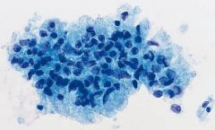
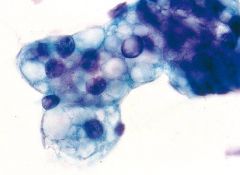
Pancreas FNA
|
Normal ductal cells
Flat cohesive sheets Even nuclear spacing, round nuclei Fine chromatin No nucleolus Distinct borders |
|
|
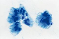
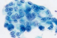
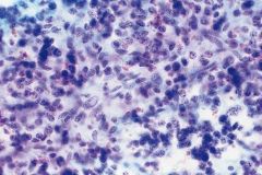
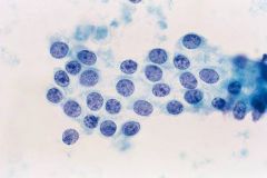
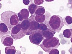
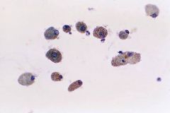
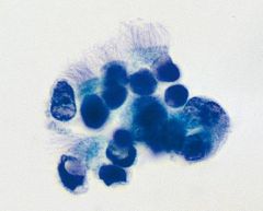
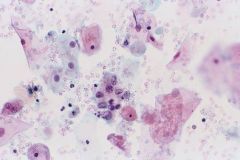
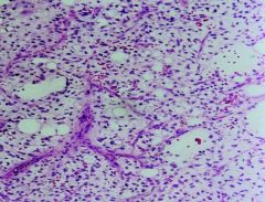
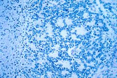
Pancreas
|
Chronic pancreatitis
Enlarged ROUND nuclei with overlapping PROMINENT NUCLEOLI Reactive conditions + for SMAD4, - p53 (vs adenoca) |
|
|
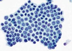
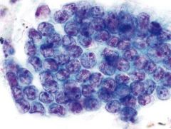
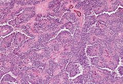
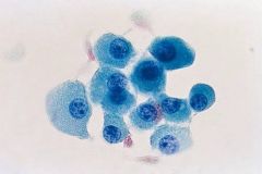
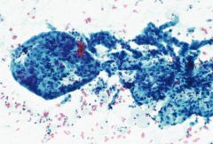
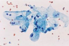
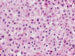
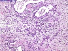
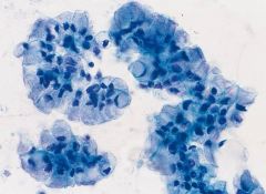
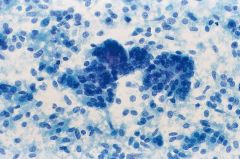
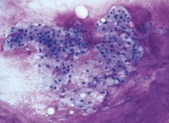
Pancreas mass head
|
Ductal adenoca
80% in head of pancreas Cellular, crowded, disordered (DRUNKEN HONEYCOMB) ISOLATED CELLS Irregular nuc contours, enlargement, irregular chromatin, mitoses ANISONUCLEOSIS (4xdifferences in size) Prominent nucleoli |
|
|
|
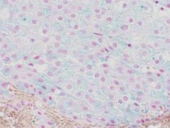
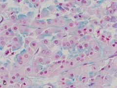
IHC of pancreatic ductal adenoca vs chronic pancreatitis vs normal GI epithelium
|
ca: +p53/-SMAD4/-CDX2
itis: -p53/+SMAD4/-CDX2 GI: -p53/+SMAD4/+CDX2 |
|
|
Pancreas
|
Adenosquamous carcinoma
Variant of ductal adeno Must have >30% squamous |
|
|
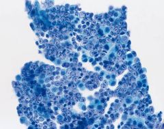
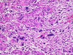
Pancreas mass
|
Undifferentiated (anaplastic) carcinoma
var of ductal adeno Highly cellular smears, pleomorphic cells Osteoclast like GIANT CELLS PHAGOCYTOSIS OF INFLAM CELLS & RBCs r/o mets |
|
|
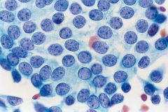
Pancreas
|
Acinar cell carcinoma
Poor prognosis Can look like benign acinar cells: usually LARGE nuclei and prominent nucleolus, with crowding. and has ISOLATED cells Can look like PEN & this is a harder distinction (both are dyshesive). Need IHC: both keratin+ ACC: trypsin, chymotrypsin, lipase PEN: chromo/synapto |
|
|
|
Acinic cell ca
Dense zymogen granules |
|
|
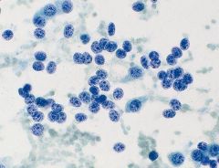
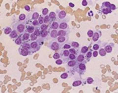
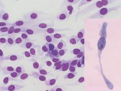
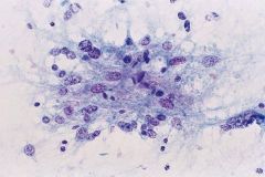
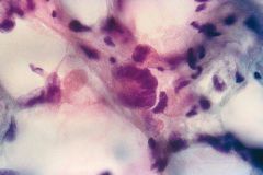
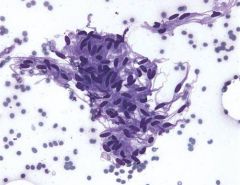
pancreas 35y F
IHC? |
Solid pseudopapillary neoplasm
Uncertain malignany potential TAIL Young women Highly cellular Hyalinized vascular stalks lined by neoplastic cells Cells not very pleomorphic; small; occasional grooves IHC? |
(+)
a1aT PR CD56 CD10 ckit. NUCLEAR BETA CATENIN+ APC gene usually negative/wk for keratins (ACC) & synapto (PEN) |
|
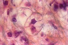
2 cm well-circumscribed pancreatic partially cystic mass
|
PEN (Pancreatic endocrine neoplasm)
Most secrete hormone (insulin, glucagon, somatostatin, VIP, serotonin, ACTH, calcitonin) Most are well-diff Predominantly ISOLATED cells, BARE NUCLEI PSEUDOROSETTES Uniform nuclei, stippled chromatin +chromo & synapto |
|
|
|
Well-diff vs poorly diff endocrine neoplasms
|
Poorly diff are uncommon; divided into 2 types:
-Small cell carcinoma -Large cell endocrine carcinoma Characterized by marked mitoses (>10/10hpf) |
|
|
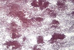
Pancreatic mass 66F
Characteristic location? |
Serous cystadenoma
BODY & TAIL Cytology: sparse cells in clean background flat sheets cuboidal cells, PAS+ glycogen in granular cytoplasm, bare nuc, small round nuc |
|
|
Pancreatic mass 50F
Location? |
Mucinous cystic neoplasm
5% pancreatic tumors Found only in women BODY & TAIL Mucinous epithelium does NOT connect to ductal system OVARIAN TYPE STROMA Can be b9, borderline, or malignant |
|
|
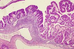
Pancreatic mass
Clinical features? Location? Most important prognostic feature? |
IPMN!
3-5% pancreatic tumors M>F! HEAD OF PANCREAS GROWS ALONG PANCREATIC DUCTS Can be b9, borderline, or malignant Most important px feature is lack of invasion (Cytology cannot distinguish MCN/IPMN) |
|
|
|
Most common mets to pancreas?
|
Lung (small cell and SCC)
Breast Kidney Lymphoma |
|
|
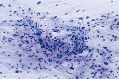
Kidney FNA
|
Normal glomerulus
-large dense globular structures with capillary loops (DDX: LG Papillary RCC but cells in gloms are NOT evenly distributed - denser in ctr - with periph capillary loops) |
|
|
Kidney
|
Proximal tubular cells
-Rare cells with round bland nucleus - Small prominent nucleolus -Abundant granular cytoplasm (granules look like they are spilling out of cytoplasm) - Lack cell borders DDx: Oncocytoma, chromophobe (but these are more cellular, frequently BINUCLEATED, variation in size, WELL DEFINED borders) |
|
|
Kidney
|
Distal tubular cells
-Rare cells, scant granular cytoplasm DDx: LG clear cell or papillary RCC (more cellular) |
|
|
Kidney FNA
IHC? |
Oncocytoma
• highly cellular • rounded nests (cell block) • cohesive fragments and dyshesive cells (smears) • abundant uniformly granular cytoplasm • Fuhrman grade 2 nucleoli r/o hepatocytes (lipofuscin) r/o RCC (more cohesive, more atypia) r/o chromophobe (less uniformly granular. Very hard to ddx! oncocytoma nested vs trabeculae of chromophobe. Hales diffuse+) EM? |
Abundant mitochondria
|
|
|
Renal cortical adenoma vs LG papillary RCC?
|
Identical except RCA < 0.5cm
|
|
|
Kidney FNA
Types? Variant? |
Angiomyolipoma
Benign PEComa Fat (scant on FNA), vessels, smooth muscle (may be atypical) 1. Young adults with TS (multiple & bilateral) 2. Middle age women (solitary) Epithelioid AML resembles ganglion cells, can have mits & necrosis IHC? |
HMB45+
MART1+ |
|
Kidney FNA 50F
|
Metanephric adenoma
rare benign Tight uniform tubules lined with bland cells with small round nuclei PSAMMOMA BODIES ~pap RCC (but are EMA-) ~WT (both WT1+ but WT is triphasic) |
|
|
Boy with renal mass, solid on imaging
|
Cystic nephroma
(aka mixed epithelial stromal tumor MEST, renatl EST = REST) Occur in boys and young women Stroma and small cysts lined by ATYPICAL EPITHELIUM (HOBNAIL) Looks solid on imaging |
|
|
mass lesion kidney
|
Xanthogranulomatous pyelonephritis
Histiocytes and MNGCs Foamy CD68+ |
|
|
|
What is the Bosniak system?
|
Classifies renal cysts by radiology
Bosniak 1: benign Bosniak 4: malignant B2&3: indeterminate |
|
|
|
What's so hard about cytology of renal cysts?
|
• Renal cysts are common, and most are benign.
• RCC can be cystic. • Adequate sampling of a cystic RCC is difficult. • Some benign cysts are difficult to distinguish from RCC: • cysts resulting from renal failure • adult polycystic kidney disease • cystic nephroma • The value of a negative diagnosis is limited. |
|
|
|
what benign entities in the kidney can have atypical cells?
|
• cystic nephroma
• renal abscess: xanthogranulomatous pyelonephritis • renal infarct • atypical cysts • angiomyolipoma Best dx clue: Hypocellular specimen |
|
|
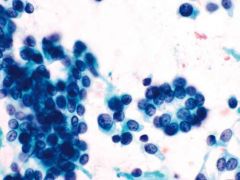
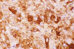
Renal mass
IHC? |
Clear cell RCC
-large cohesive cell groups with transgressing vessels -Abundant wispy cytoplasm with ILL-DEFINED edges -Vacuoles -Large round eccentric nuc CAN HAVE BASEMENT MATERIAL BLOODY +CD10, vimentin, RCC genetics? |
3p-
(VHL gene) |
|
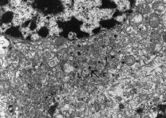
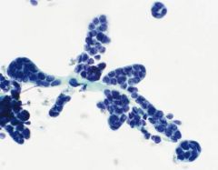
renal mass
|
Papillary RCC (10% RCCs)
Type 1: Fuhrman1,2. Small, scant cyt. Low grade, more common. foamy macrophages and i/cyt hemosiderin Type 2: Large cells abundant granular cyt. Large nuc, prom nucleoli (F3) Note large sphere in 1st image, characteristic IHC? |
+EMA, LMWK, CK7
-34BE12, WT1 |
|
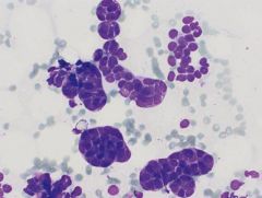
Renal mass
Genetics? |
Papillary RCC
Hemosiderin in cyt Note foamy macs stuffing papillary cores Trisomy 7, 16, 17 |
|
|
Hales colloidal iron. Tumor?
|
CHROMOPHOBE has diffuse staining. What does oncocytoma look like?
|
Luminal staining only.
|
|
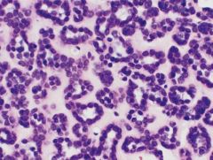
Kidney mass
|
Chromophobe
3-5% RCC Trabeculae with abundant cytoplasm, DISTINCT BORDERS, dark chromatin, RAISINOID nuc outlines (look like koilocytes...) DDx: Clear cell RCC, oncocytoma Genetics? |
Monosomies
|
|
|
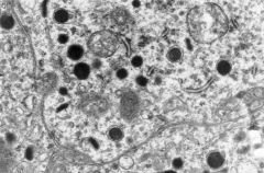
Chromophobe
polygonal tumor cells with abundant granular cytoplasm, round nuclei and well-defined cytoplasmic membrane (pic) perinuclear vacuolization & multinucleation EM? |
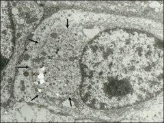
Numerous microvesicles pushing the nucleus
|
|
Renal mass
|
Sarcomatoid rcc
high grade spindle cell neoplasm poor px |
|
|
Medullary mass
|
Collecting duct carcinoma of bellini
Rare MEDULLARY Tubulopapillary histology High grade cytology (like a met) PROMINENT DESMOPLASIA ***++34Be12 |
|
|
|
What should you think about with a renal mass in a child that resembles RCC?
|
Translocation-associated renal cell carcinoma
Chromosome X Transcription factor E3 gene (TFE3) (IHC TFE+, EMA weak) Ca++ |
|
|
Renal pelvic mass
|
Urothelial carcinoma
Large cells with dark nuclei. Dense smooth cytoplasm. ELONGATED cells "Cercariform" (tadpole) cells IHC? |
UCC: + 34Be12, CK20, CEA, mucin
RCC: CD10, RCC, PAX2 |
|
|
Most common met to the kidney?
|
Lung
|
|
|
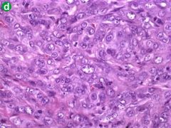
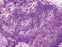
Adrenal mass
|
Myelolipoma
Fat and hematopoietic elements |
|
|
Solitary adrenal mass
|
Adrenal cortical adenoma
Common 85% on-functioning (vs carcinomas - most are fxnl) Numerous NAKED nuclei in a frothy background (<3.5cm "benign cortical nodule") |
|
|
Adrenal mass >5cm
|
Adrenal cortical carcinoma
Uncommon Usually fxnl usually >5cm Isolated cells with INTACT granular cytoplasm (vs adenoma) Pleomorphic nuclei Mitoses IHC? |
inhibin+
Calretinin+ MelanA+ (-)EMA, CEA, keratin |
|
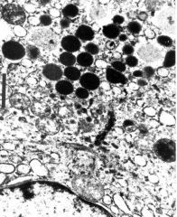
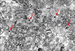
Adrenal mass
EM? |
Pheochromocytoma
EM shows neurosecretory granules (dark balls) Adrenal medulla HTN 20% assoc with familial syndromes (which ones?) 10% bilateral DO NOT FNA! cellular, loose clusters and isolated cells. Pleomorphic cells, stippled chromatin, INCIs*** |
MEN2a
MEN2b Neurofibromatosis VHL |
|
|
Most common mets to adrenal?
|
Lung
RCC Melanoma |
|
|
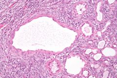
Ovarian cyst
|
Benign follicular cyst
coarsely granular chromatin • mix of viable and pyknotic nuclei • foamy cytoplasm • mitoses High estradiol levels (E2) |
|
|
Ovarian cyst
|
Corpus luteal cyst
Isolated luteinized granulosa cells with abundant finely vacuolated cyt |
|
|
Ovarian cyst
|
Endometriotic cyst
|
|
|
ovarian cyst
|
detached cilia
exclude follicular cyst seen in serous cyst, hydrosalpinx, cystic teratoma |
|
|
Ovarian cyst
|
Serous cystadenoma
ELEVATED CA125 in cyst fluid |
|
|
ovarian cyst
Fluid chemistry? |
mucinous cystadenoma
HIGH CEA LOW CA-125, E2 |
|
|
ovarian cyst
|
serous borderline tumor
atypia Psammoma bodies |
|
|
Ovarian cyst
|
Serous cystadenocarcinoma
Marked nuclear atypia |
|
|
ovarian cyst, large multiloculated
|
mucinous cystadenocarcinoma
|
|
|
Ovarian cyst
|
Endometrioid adenocarcinoma
|
|
|
Ovarian cyst
|
Cystic teratoma
|
|
|
Ovarian mass
|
Granulosa cell tumor
Similar to benign granulosa cells... Adult type: 95%, postmenopausal. secrete estrogen highly cellular. Call-Exner bodies. Rd nuc with grooves. Juvenile type: kids & teens. secrete estrogen. solid. LACKS NUC GROOVES. only 10% aggressive despite high mits. r/o small cell ca of ovary (same age) IHC? |
ALPHA-inhibin
CD99 calretinin S100 punctate cytokeratin SMA (-)CK7, EMA |
|
|
Most common mets to ovary?
|
GU
Colon Stomach (Krukenberg tumor) Breast |
|
|
soft tissue
|
fat necrosis
lacks scalloped nuc of lipoblasts |
|
|
soft tissue
|
Floret cell of pleomorphic lipoma
|
|
|
|
Lipoblast
hyperchromatic, scalloped nuc, large vacuoles |
|
|
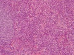
soft tissue
Extremity 70y.o. |
Myxoid MFH/ myxofibrosarcoma
Curvilinear blood vessels Marked peomorphism |
|
|
deep thigh mass
|

low-grade fibromyxoid sarcoma
looks bland but 30% metastasize myxoid matrix bland spindle cells genetics? |
t(7;16)
|
|
40y.o. thigh mass
|
Myxoid liposarcoma
CHICKEN WIRE VESSELS SIGNET RING LIPOBLASTS microcystic change Sheets of small uniform cells Genetics? Unique mets? |
t(12;16)
mets to SOFT TISSUE > spine > lung |
|
Axial skeletal mass
|
Chordoma
• granular and fibrillary myxoid matrix • cohesive clusters and cords of neoplastic cells • comparatively large cells PHYSALIPHOROUS CELLS! (bubbly) IHC? |
cytokeratin AND S100 positive
|
|
40y.o. thigh mass
|
Extraskeletal myxoid chondrosarcoma
distinctive fibrillar chondromyxoid matrix IHC? genetics? unique mets? |
<20% S100
t(9;22) EWS gene LATE mets (10y) |
|
|
Schwannoma
• large, cohesive fragments • wavy, “fishhook” nuclei • pointed nuclear ends • nuclear palisading • filamentous cytoplasm S100+ |
|
|
Teenager thigh mass
|
Synovial sarcoma
Characteristic pattern of cohesive cell clusters alternating with dispersed cells genetics? |
t(x;18)
|
|
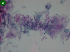
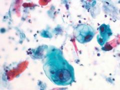
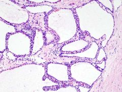
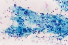
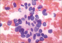
Pancreatic cyst, superficial, multilocular
|
Lymphoepithelial cyst
Middle age men Nuc & anucleated squames lymphs (but squames dominate) |
|
|
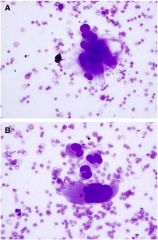
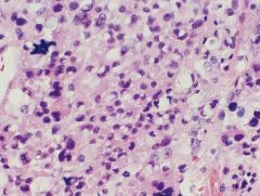
1y.o. with adrenal mass
IHC? |
Neuroblastoma
Small primitive cells Homer-Wright rosettes Neuropil IHC: (+)Chromo, synapto, ALK EM? |
NEUROSECRETORY GRANULES
|
|
Thigh 19F
IHC? genetics? |
Ewings sarcoma /PNET
CD99+, PAS+ t(11;22) What else stains with CD99? small uniform cells with rosettes, pseudorosettes Cytoplasmic vacuoles |
Synovial sarcoma
Embryonal RMS |
|
|
Characteristics of alveolar rhabdomyosarcoma?
|
round cell tumor extremities young patients. Highly cellular. Irreg nuclei.
Myogenin+ t(2;13), t(1;13) |
|
|
Thigh young woman
|
Alveolar soft part sarcoma
Bloody smears Clusters of cells with round to oval nuclei, prominent large nucleoli, abundant pale finely vacuolated to granular cytoplasm, with fraying of the cytoplasmic margins. PAS+ 1/cyt magenta pink granules and occasional crystals. |
|
|
|
Renal mass child
|
Wilms tumor
TRIPHASIC (epith, stroma, blastema) Blastema cells are small round blue cells, can form rosettes |
|