![]()
![]()
![]()
Use LEFT and RIGHT arrow keys to navigate between flashcards;
Use UP and DOWN arrow keys to flip the card;
H to show hint;
A reads text to speech;
35 Cards in this Set
- Front
- Back
|
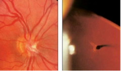
Pseduopapilledema: Glial remnant on optic disc, persistant hyaloid tissue (Bergmeister's papilla) |
|
|
Morning glory coloboma of disc |
|
|
Optic disc of hypoplasia |
|
|
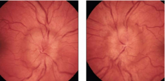
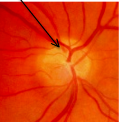
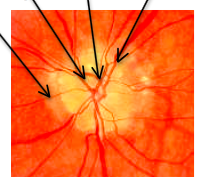
Early papilledema -- blurring (swelling) of peripapillary NFL, blurring of disc margins (inferior-superior poles)
|
|
|
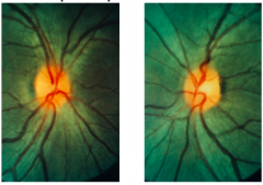
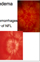
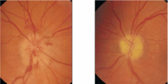
Early papilledema -- splinter flame-shaped NFL hemorrhage (left), NFL swelling (right), disc hyperemia, optic nerve cupping retained
|
|
|
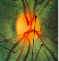
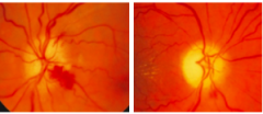
Papilledema -- both disc margins are obscurred and nerve is becoming elevated due to swelling. Can also see hemorrhage at 5 o'clock |
|
|
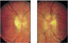
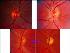
Stages of papilledema -- venous engorgement, disc hyperemia (dilation of disc capillaries) |
|
|
Drusen of optic nerve |
|
|
Drusen of optic nerve |
|
|
Swollen disc |
|
|
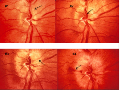
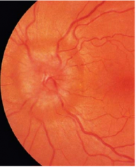
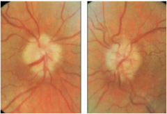
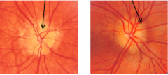
Moderate papilledema -- increased disc hyperemia, elevation of dic surface, obscuration of BVs crossing disc surface, thickening & opacification of NFL, venous engorgement & increased tortuosity
|
|
|
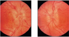
Moderate papilledema -- Optic nerve cupping lost, Paton's lines appear as circumferential "wrinkles" adjacent to the disc
|
|
|
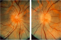
Chronic papilledema |
|
|
Chronic papilledema |
|
|
Atrophic papilledema – chronic disc edema |
|
|
Atrophic papilledema -- resolving edema |
|
|
Atrophic papilledema with disc pallor |
|
|
Atrophic papilledema |
|
|
Papillitis OD |
|
|
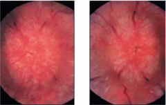
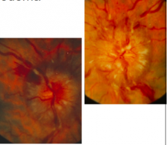
Fully developed papilledema -- lipid exudates, marked disc elevation, increased disc hyperemia, obscuration of BVs crossing disc surface, venous engorgement & increased tortuosity, CWS, retinal hemorrhages
|
|
|
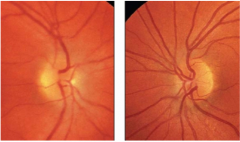
Chronic papilledema -- less disc hyperemia; decrease in edema, CWS, hemorrhages; less NFL swelling as atrophy of NFL begins; without viable NF there is less axoplasmic obstruction so less clinical presentation |
|
|
Atropic papilledema -- marked reduction in edema, hemorrhages; sheathing of retinal vessels; pallor of optic nerve
|
|
|
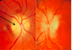
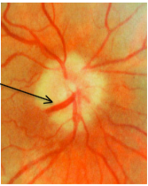
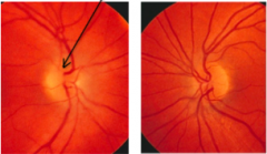
Atropic papilledema -- optociliary shunt vessel formation; retinochoroidal venous collateral vessels bypass region of obstruction.
|
|
|
Optic neuritis (papillitis) -- unilateral optic nerve edema |
|
|
Optic neuritis (papillitis) -- right optic nerve is edematous, white and smudged borders, disc margin is obscured. Will present with APD and color desaturation |
|
|
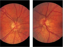
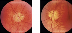
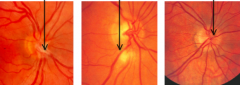
Anterior ischemic optic neuropathy (AION) -- left: Acute AION -- unilateral nerve edema, disc hemorrhage common right: old "inactive" AION -- optic nerve atrophy with pallor |
|
|
Non-arteritic (NAION) -- optic nerves small in diameter with small cupping |
|
|
AION OD, optic nerve pallor & atrophy OS from previous AION episode |
|
|
AION & NAION -- disc hemorrhage (left) & unilateral optic nerve pale edema - sectoral (right) |
|
|
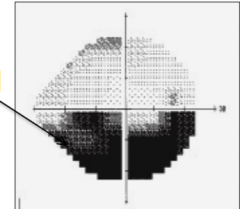
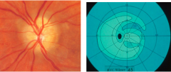
Altitudinal VF defect in AION and NAION |
|
|
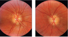
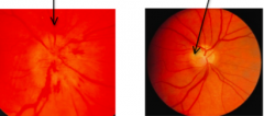
Pseudopapilledema -- optic nerve appearance simulates disc edema
left: persistant hyaloid tissue (Bergmeister's papilla) middle: medullated nerve fibers right: hyperopia - small optic nerves, nerve hypoplasia |
|
|
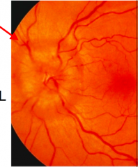
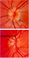
Pseduopapilledema -- drusen of the optic nerve; yellowish glistening spheres on or within the optic nerve. Nerves appear elevated "full" or "swollen" |
|
|
Pseduopapilledema -- drusen of the optic nerve; left: peripapillary region RPE irregular middle left: retinal vessels exit nerve centrally middle right: retinal vessels anomalous branching patterns right: retinal vessels clearly visible crossing disc surface |
|
|
Pseduopapilledema -- drusen of the optic nerve; disc margins poorly defined, no or very small physiological cup, VF defects: progressive & varied |
|
|
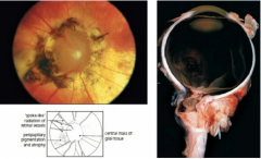
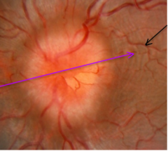
Drusen of the optic nerve: 1) retinal vessels: exit nerve centrally, clearly visible crossing disc surface, anomalous branching 2) disc color WNL or slightly hyperemic 3) peripapillary RPE irregular 4) disc margins poorly defined 5) no or very small physiological cup 6) disc appears slightly "full" "swollen" 7) yellowish glistening spheres 8) no hemorrhage 9) no venous engorgement 10) no CWS |