Reading...
![]()
Play button
![]()
Play button
![]()
Use LEFT and RIGHT arrow keys to navigate between flashcards;
Use UP and DOWN arrow keys to flip the card;
H to show hint;
A reads text to speech;
49 Cards in this Set
- Front
- Back
|
Where are B cells typically found in lymph nodes?
|

Cortex
|
|
|
Where are T cells typically found in lymph nodes?
|
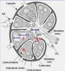
Paracortex
|
|
|
Where do lymphomas originate from in the lymph nodes?
|

The germinal centers
|
|
|
Where do mantle cell lymphomas originate from?
|
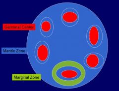
the mantle zone
|
|
|
What is a cause of marginal zone expansion in lymph nodes?
|

Hyperplastic syndromes
Marginal zone lymphomas |
|
|
What is a primary follicle?
|
A follicle without a germinal center
|
|
|
What are the most common types of B-cell non-Hodgkins lymphoma?
|
1. Diffuse large B cell lymphoma
2. Follicular lymphoma |
|
|
What are the most common types of Hodgkin lymphomas?
|
Classic hodgkin lymphoma
|
|
|
What are the different types of B-cell non-Hodgkin lymphomas?
|
Chronic lymphocytic leukemia/small lymphocytic lymphoma
Follicular lymphoma Mantle cell lymphoma Diffuse large B-cell lymphoma Marginal zone lymphoma |
|
|
What are the aggressive lymphomas?
|
Mantle cell lymphoma
DLBCL Burkett lymphoma |
|
|
What are the more indolent lymphomas?
|
CLL/SLL
Marginal zone Follicular |
|
|
What does it mean when referring to a B-cell lymphoma that is restricted?
|
Monoclonal expression of a kappa;lambda light chain as determined by flow cytometry
This is abnormal! |
|
|
What are good markers for B cells?
|
CD20
|
|
|
What are good markers for T cells? What do you have to confirm?
|
CD2, 3, 5, 7
You need to see that they're expressed at the same level to confirm that there's not a T cell lymphoma |
|
|
What does CLL/SLL look like?
|
Nuclei that are odd looking
Too many T cells |
|
|
How do you confirm a diagnois of CLL/SLL?
|
Flow cytometry
Biopsy |
|
|
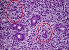
What's diagnostic for CLL/SLL in a lymph node?
|
Proliferation centers!
lots and lots of the CLL/SLL cells all growing in one place |
|
|
What immunohistochemistry stains are used to diagnose CLL/SLL?
|
CD20
CD5 CD5 is aberrantly expressed...normally it's a T cell marker! |
|
|
What do you see in flow cytometry for CLL/SLL?
|
Dim CD20
Dim immunoglobulin +CD23 |
|
|
Where can mantle cell lymphomas present?
|
Nodes
Inside the intestine as polyps Spleen |
|
|
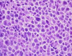
What is the appearance of the cells in mantle cell lymphomas?
|
More angular
Lots of them Irregular nuclear patterns |
|
|
What marker is specific for mantle cell lymphoma?
|
Cyclin D1
|
|
|
What are the immunohistochemial findings in mantle cell lymphoma?
|
+CD20
+CD5 +cyclin D1 |
|
|
What do you find in flow cytometry for mantle cell lymphoma?
|
Moderate to bright CD20
Moderate to bright light chain |
|
|
What do you find on FISH for mantle cell lymphoma?
|
t(11;14) CCND1/IgH
|
|
|
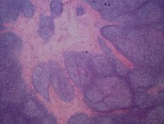
What does a lymph node look like in follicular lymphoma?
|

Nodularity!
Lack of normal node structure |
|
|
What is the appearance of the cells in follicular lymphoma?
|
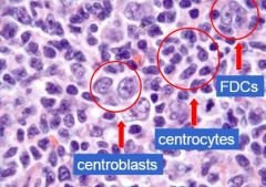
Small cells
Small cells with clefts: centrocytes Hollowed out nuclei with multiple nucleoli: centroblasts (grading) Dispersed chromatin with a central nucleoli: FDC (follicular dendritic cells) |
|
|
What makes a follicular lymphoma higher grade?
|
More centroblasts per high powered field.
|
|
|
What are the findings in immunohistochemistry for follicular lymphomas?
|
CD20
Germinal center markers: CD10 BCL6 |
|
|
What do you find on FISH in follicular lymphoma?
|
t(14,18): immunoglobulin heavy with the BCL2
|
|
|
What's the zoomed out appearance of marginal zone lymphoma?
|
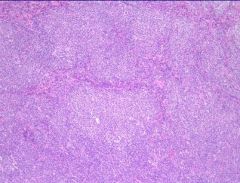
Monocytoid: it looks more white than normal
Colonization of the germinal centers |
|
|
What is a Dutcher body? What condition is it found in?
|
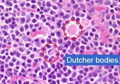
Intranuclear inclusions of IgGs
It's found in marginal zone lymphomas |
|
|
What are the subtypes of marginal zone lymphomas?
|
Extranodal MALT: stomach, conjuctiva, salivary gland
Nodal marginal zone lymphoma Splenic marginal zone lymphoma |
|
|
What is a common cause of gastric MALTs?
|
H. pylori infection
|
|
|
What do you see in marginal lymphomas?
|
Lymphoepithelial lesions: the lymphocytes are invading the epithelium!
|
|
|
What's the problem with the diagnosis of marginal zone lymphomas?
|
They don' have any specific immunohistochemical markers
You can only use CD20 to confirm it's a B-cell; it's a diagnosis of exclusion |
|
|
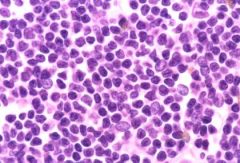
What's the appearance of the cells in a diffuse large B-cell lymphoma?
|
Big!
Dark/hyperchromatic Atypical looking! |
|
|
What do you find in large b cell lymphoma more than in other types?
|
Necrosis.
|
|
|
What immunohistochemistries do you do for large B cell lymphomas?
|
CD20: see if it's a B cell
10: see if it came from a germinal center |
|
|
What's the progression of a Burkitt lymphoma?
|
Fast; it's an aggressive mass
|
|
|
How does a Burkitt lymphoma typically present?
|
Terminal mass in the ileum of a child
|
|
|
What are the types of hodgkin lymphoma?
|
Classical hodgkin lymphoma
Nodular lymphocyte predominant hodgkin lymphoma |
|
|
What are the subtypes of classical hodgkin lymphoma subtypes?
|
Nodular sclerosis
Mixed celluarity (inflammatory cells) Lymphocyte rich (lymphocytes all over the place) Lymphocyte depleted (rare!) |
|
|
How does nodular sclerois typically present?
|
Mediastinal mass in a young person
|
|
|
What are the type of neoplastic cells in the nocular sclerosis subtype of hodgkin lymphoma?
|
Reed-sternberg cells/variants
|
|
|
What is the typical appearance of Reed-Sternberg cells?
|
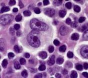
Lots of free space
Binucleate Prominent nuclei They look like owl eyes! |
|
|
What type of cells are reed-sternberg cells?
|
B cells
|
|
|
What do you almost always see in Hodgkin lymphomas?
|
Inflammatory background
|
|
|
What is the immunophenotype for Hodgkin lymphoma?
|
CD30
CD15 |