![]()
![]()
![]()
Use LEFT and RIGHT arrow keys to navigate between flashcards;
Use UP and DOWN arrow keys to flip the card;
H to show hint;
A reads text to speech;
74 Cards in this Set
- Front
- Back
|
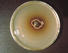
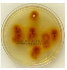
black, yeasty colony. covering of short olive gray mycelium
|
Hortaea werneckii
|
|
|
Will not grow on routine media unless layered with olive oil (or lipid) and incubated at 37 C
|
Malassezia furfur
|
|
|
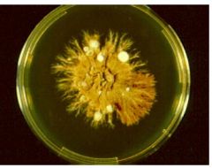
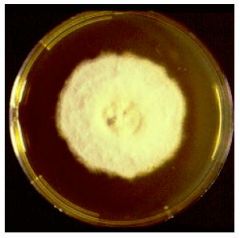
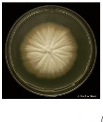
Slow growth Dark green to black heaped glabrous colony |
Piedraia hortae |
|
|
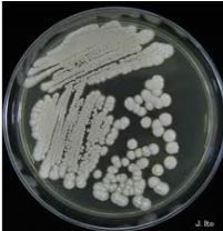
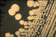
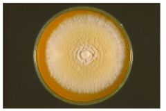
Cream, wrinkled, glabrous colony |
Trichosporon beigelii
|
|
|
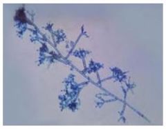
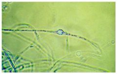
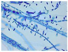
Yeast portion contains dark one or two celled blastoconidia. The mold portion also contains annellides with clusters or chains of one or two celled dark annelloconidia |
Hortaea werneckii |
|
|
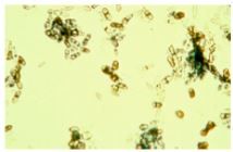
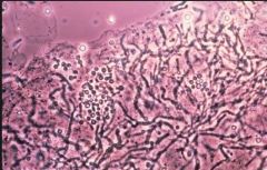
“Spaghetti and meatballs” Thick round to oval cells in clusters with short, angular hyphae; budding yeast with septate |
Malassezia furfur |
|
|
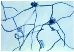
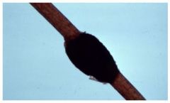
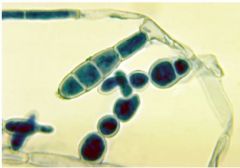
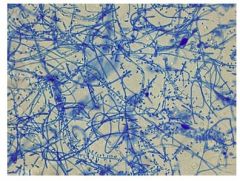
Dark thick - walled hyphae with swellings. Asci and Ascospores may be present. |
Piedraia hortae |
|
|
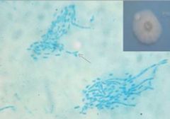
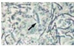
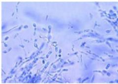
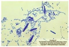
Blastoconidia and Arthroconidia |
Trichosporon beigelii |
|
|
Tinea nigra
Brown to black non-scaly patches form primarily on the palms of the hands and feet. |
Hortaea werneckii
|
|
|
pityriasis versicolor, tinea versicolor. scaly patches of different colors that fluoresce under Wood’s lamp. Can cause systemic disease
|
Malassezia furfur
|
|
|
Black Piedra. black nodules around scalp hairs
|
Piedraia hortae
|
|
|
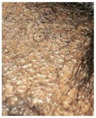
White Piedra. Light brown soft
nodules around beard and mustache hairs |
Trichosporon beigelii
|
|
|
Hyphal elements fragment at septation points. look box -like.
|
Arthroconidia
|
|
|
Worldwide distribution –More prevalent in hot, humid |
Malassezia furfur |
|
|
Endemic in tropical regions of Africa, Asia and Latin America |
Piedraie hortae |
|
|
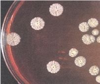
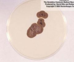
Grows slowly on Sabouraud dextose agar at Room Temperature •Forms brown restricted colonies |
Piedraie hortae |
|
|
Piedraie hortae |
|
|
Most pathogenic Trichosporon species |
T. asahaii |
|
|
Widely distributed in topical areas |
Trichosporon species |
|
|
Absence of carbohydrate fermentation, Urease positive |
Trichosporon species |
|
|
Trichosporon species |
|
|
Trichosporon species |
|
|
Trichosporon species |
|
|
Endemic in tropical areas of Central and South America, Africa and Asia |
Hortaea werneckii |
|
|
Hortaea werneckii |
|
|
Hortaea werneckii |
|
|
Hortaea werneckii |
|
|
Malasazzia furfur |
|
|
Piedra hortae |
|
|
Piedra hortae |
|
|
Trichosporon beigelli |
|
|
Malassezia furfur |
|
|
Malassezia furfur |
|
|
Scatula |
Cup shaped, crusty flakes |
|
|
Tinea favosa or favus |
Trichophyton schoenleinii infection of hair follicle that progresses to a crusty lesion of dead cells and fungal mycelia |
|
|
Tinea capitis |
Gray-patch ringworm & Black-dot ringworm |
|
|
Common childhood disease |
Gray-patch ringworm |
|
|
Endothrix hair involvement |
Black-dot ringworm |
|
|
Black dot ringworm |
|
|
Onychomycosis |
Nail Infections |
|
|
common causes of Onychomycosis |
Trichophyton tubrum |
|
|
best treatments for Onychomycosis |
Terbinafine and itraconazole |
|
|
Tinea pedis |
Athlete's Foot |
|
|
Systemic Dermatophyte Infections |
Granulomas –May have spread from athlete’s foot or fungal infection of nails |
|
|
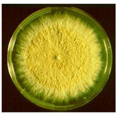
Epidermophyton floccosum |
|
|
Epidermophyton floccosum |
|
|
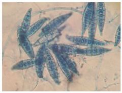
Microsporum canis |
|
|
Microsporum canis |
|
|
Microsporum gypseum |
|
|
Microsporum gypseum |
|
|
Microsporum audouinii |
|
|
Microsporum audouinii |
|
|
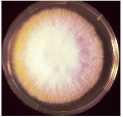
Trichophyton mentagrophytes |
|
|
Trichophyton mentagrophytes |
|
|
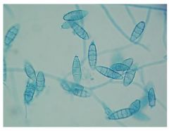
Trichophyton rubrum |
|
|
Trichophyton rubrum |
|
|
Trichophyton tonsurans |
|
|
Trichophyton tonsurans |
|
|
Direct examination of the sample is performed with |
10% KOH |
|
|
begins at the root of the hair, moves through the hair shaft and is displayed externally further up the hair |
Endothrix invasion |
|
|
Ectothrix infections are caused by |
Microsporum and Trichophyton mentagrophytes, rubrum and verrucosum. |
|
|
Endothrix hair infections are only caused by |
Trichophyton tonsurans, schoenleinii, and violaceum. |
|
|
examination with wood’s lamp cannot rule out fungal infections. |
all fungi that infect hairs will produce this fluorescent metabolite; |
|
|
Thick walled hyphal element produced during unfavorable growth conditions that will develop conidia when conditions are better |
Chlamydoconidia |
|
|
Fungus that affects the dermis (hair, skin, or nails) and belongs to Epidermophyton, Microsporum, or Trichophyton genera. |
dermatophyte |
|
|
Infection around the outside of the hair shaft. The infection starts in the root inside the shaft, but the arthroconidia develop around the hair further up the shaft. The cuticle is destroyed by this type of infection. |
ecothrix |
|
|
Infection with arthroconidia inside the hair shaft. The infection starts in the root, travels up the hair shaft and may appear on the outside of the hair somewhere higher up the shaft. The cuticle is not destroyed
|
endothrix
|
|
|
Blunt branched hyphae that resemble the horns of a buck deer (if you have a vivid imagination). Predominant feature in T. schoenleinii |
Favic chandeliers |
|
|
knot of twisted hyphae, often seen in older cultures |
nodular bodies |
|
|
hyphae resembling tennis racquets placed end to end |
raquet hyphae |
|
|
hyphae presenting as a spiral, either flat or three dimensional |
spiral hyphae |
|
|
Causes over 90% of the dermatophytosis of the scalp in the US today. Epidemic in black school age children. |
T. tonsurans |
|
|
The hair perforation test is used to differentiate |
Trichophyton mentagrophytes from Trichophyton rubrum- Hair will be perforated by T. mentagrophytes and not by T. rubrum |
|
|
Most common cause of athlete’s foot, also infects body, nails, beard and scalp (Exothrix). |
T. mentagrophytes |