Reading...
![]()
Play button
![]()
Play button
![]()
Use LEFT and RIGHT arrow keys to navigate between flashcards;
Use UP and DOWN arrow keys to flip the card;
H to show hint;
A reads text to speech;
53 Cards in this Set
- Front
- Back
|
What is Monday's quiz over?
|
4/15 and 4/26
|
|
|
What is the flank fold?
|
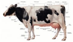
The area that horses and bovids don't like to be kicked in formed of cutaneous trunci m. and skin.
|
|
|
How many lumbar vertebrae do horses have?
|
Usually 6 lumbar vertebrae present, although 5 have been reported in the domestic horse, donkey, Arabian horse, Przewalski horse, ass, and mule
|
|
|
Describe the transverse processes of the lumbar vertebrae of the horse.
|
Length increases to the third or fourth then decreases to the last
First two curve caudally, last two curve cranially L5 articulates with L6; L6 articulates with sacrum |
|
|
How many lumbar vertebrae do ox have? How do they differ from the horse?
Describe their transverse processes. |
Usually 6 lumbar vertebrae present
Lumbar vertebrae are much longer than in the horse Articular processes are large and their facets are more curved than in the horse Transverse processes: All curve cranially Shortest on L1; length increases to L5; shorter on L6 |
|
|
What is the area between the last rib and the tuber coxae of the ox?
What is the clinical relevance of this region? |
The paralumbar fossa.
Boundaries of the paralumbar fossa Last rib Tuber coxae Transverse processes of the lumbar vertebrae This is the region where the incision is made in standing surgeries of ox. |
|
|
Do horses tolerate standing abdominal surgeries?
|
Usually not - but they do general anesthesia much better than do ox.
|
|
|
What are the lymph nodes just cranial to the horse and ox thigh? What are they located between? Where do they drain to?
|
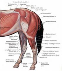
Subiliac lymph nodes in the horse
Also known as prefemoral lnn. Horse: Located craniomedial to the tensor fasciae latae m., halfway between the tuber coxae and patella Ox: Located on the aponeurosis of the external abdominal oblique m. close to tensor fasciae latae m. Efferents follow the ventral brs. of the deep circumflex vessels to the lateral and medial iliac lymph nodes |
|
|
What nerves form the lateral cutaneous femoral n.? What does this nerve innervate?
|
Lateral cutaneous femoral n. (formed from ventral brs. of L3, L4 +/- L5 spinal nn.)
Innervates skin in the cranial, medial thigh region |
|
|
What assists the abdominal muscles in supporting the weight of the abdominal viscera?
What does this structure cover laterally and ventrally? |

Tunica flava abdominis a deep fascial sheet of elastic tissue.
Ventrally it covers and adheres to the aponeurosis of the external abdominal oblique m. Laterally, it covers the external abdominal oblique, external intercostal, and serratus ventralis thoracis mm. |
|
|
What is the median fibrous raphe extending from the xiphoid cartilage to the prepubic tendon?
What is this formed from? |
Linea alba
Formed by the junction of aponeuroses of external abdominal, internal abdominal and transversus abdominis mm. |
|
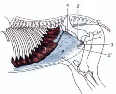
What are these structures?
|

2 aponeurosis
2' pelvic tendon 2" abdominal tendon 3 superficial inguinal ring 4 heave line |
|
|
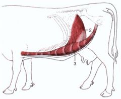
What are the OIAI of the external abdominal oblique m.?
Which ways do the fibers run? |

O: Lateral surfaces of 4th-18th ribs and the fascia over the external intercostal mm.; thoracolumbar fascia
I: Linea alba and prepubic tendon, tuber coxae and body of the ilium, medial femoral fascia Innervation: Ventral brs. of thoracic and lumbar spinal nn. Action: Compress the abdominal viscera, as in defecation, urination, parturition and expiration; flex the trunk Fibers directed ventrocaudally |
|
|
What are the OIAI of internal abdominal oblique m.? Which direction do the fibers course?
|
O: Tuber coxae and adjacent part of the inguinal ligament
I: Cartilages of the last 4-5 ribs; linea alba and prepubic tendon Innervation: Ventral brs. of thoracic and lumbar spinal nn. Action: Compress the abdominal viscera, as in defecation, urination, parturition and expiration; flex the trunk Fibers course ventrally, cranially and medially |
|
|
What are the OIAI of the transversus abdominis m. of the horse?
What is different in the ox? |
O: Medial surface of the ventral ends or cartilages of the asternal ribs and the transverse processes of the lumbar vertebrae via thoracolumbar fascia
I: Xiphoid cartilage and linea alba Innervation: Ventral brs. of thoracic and lumbar spinal nn. Action: Compress the abdominal viscera, as in defecation, urination, parturition and expiration; flex the trunk Dorsal part is muscular, ventral part is aponeurotic. In the ox it goes aponeurosis, muscle, aponeurosis. |
|
|
What are the OIAI of the rectus abdominis m?
|
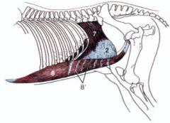
8
O: Cartilages of the 4th or 5th to 9th ribs and the adjacent surface of the sternum I: Pubis via the prepubic tendon Innervation: Ventral brs. of thoracic and lumbar spinal nn. Action: Similar to that of external and internal abdominal oblique mm. (abdominal compression); flex the lumbosacral joints and the lumbar and thoracic parts of the spine |
|
|
What are the OIAI of the transversus abdominis m in the ox?
|
O: Deep lumbar fascia from the transverse processes of the first 5 lumbar vertebrae, medial surface of the ventral ends or cartilages of the asternal ribs, the transversalis fascia, and the caudomedial surface of the last rib
I: Linea alba Innervation: Ventral brs. of thoracic and lumbar spinal nn. Action: Retract the ribs and compress the abdominal viscera Dorsal part is aponeurotic, costal attachment is muscular |
|
|
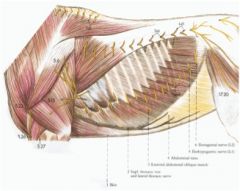
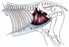
What layers of muscles will you be incising through when you cut into the paralumbar fossa?
|
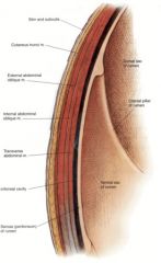
see the picture
|
|
|
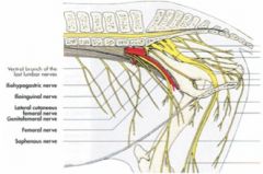
How far will the dorsal branches of the spinal nerves course?
|
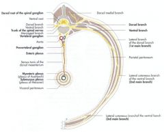
Extends ventrally to the level of the stifle or patella - so you need to block both the ventral and dorsal branches when working in this area.
Look at the cutaneous branches here. |
|
|
What nerves innervate the flank of the ruminant?
|
Dorsal and ventral brs. of T13, L1, L2, L3 spinal nerves
Ventral brs. T13 – costoabdominal n. L1 – iliohypogastric n. L2 – ilioinguinal n. L3 – genitofemoral n. |
|
|
To perform surgery in the paralumbar region, what nerves needs to be anesthetized?
|
To perform surgery in the paralumbar region, it is necessary to anesthetize the dorsal and ventral branches of T13, L1 and L2 spinal nerves
|
|
|
What are the boundaries to the paravertebral block of the paralumbar region of the ruminant?
|
You only perform this block on the side that you are performing surgery.
T13 spinal n. Palpate last rib and transverse processes of L1 and L2 Insert needle vertically 5 cm in ox (3 cm in sheep and goat) from dorsal midline in a transverse plane at the cranial angles of the tips of the transverse process of L1. Anesthetize both dorsal and ventral branches of T13 since they are close together at this point. L1 and L2 spinal nn. Make similar injections at the transverse planes at the caudal borders of the transverse processes of L1 and L2 Nerves are infiltrated close to the vertebrae |
|
|
What are the advantages and disadvantages to the paravertebral block of the paralumbar fossa of the ruminant?
|
Nerves are infiltrated close to the vertebrae
Advantages: Uniform anesthesia of all structures of the paralumbar fossa including the peritoneum Good muscle relaxation Disadvantages: Technique important to avoid puncturing the aorta, caudal vena cava or azygous v. Paralyzes back muscles on the side of the operation causing a convexity of the trunk Viscera may bulge out of the incision Closure is more difficult |
|
|
What are the boundaries to the paralumbar block of the paralumbar region of the ruminant?
|
You only perform this block on the side that you are performing surgery.
T13 spinal n. Dorsal and ventral to the tip of L1 transverse process and between the last rib and transverse process L1 spinal n. Dorsal and ventral to the caudal edge of L2 transverse process L2 spinal n. Dorsal and ventral to the caudal edge of L4 transverse process May need additional injection midway between L3 and L4 vertebrae since we're performing this laterally. Nerves are blocked at the tips of the transverse processes |
|
|
What are the advantages and disadvantages to the paralumbar block of the paralumbar fossa of the ruminant?
|
Nerves are blocked at the tips of the transverse processes
Advantages: Uniform anesthesia of all structures of the paralumbar fossa including the peritoneum Good muscle relaxation Back muscles are not paralyzed Disadvantages: Possible individual variation in position of spinal nerves may necessitate additional injections |
|
|
What are the nerves you'd block and the site of injection of the lumbar epidural in the ruminant?
|
Block T13, L1, L2 spinal nerves
Site of injection – between L1 and L2 vertebrae |
|
|
What nerves need to be blocked to anesthetize the udder of the cow, ewe and doe?
|
Cow - block ventral brs of L1-L4 spinal nerves
Ewe and doe - block ventral brs of L2-L4 spinal nerves |
|
|
Where does the costal part of the horse diaphragm attach?
|
Attaches to the cartilages of ribs 8-10, ribs 11-18
|
|
|
Where does the sternal part of the horse diaphragm attach?
Where do the lumbar parts of the diaphragm of the horse attach? |

Dorsal surface of xiphoid cartilage
Right crus – lumbar vertebrae 1-4 (or 5) via ventral longitudinal ligament Left crus – lumbar vertebrae 1 and 2 via ventral longitudinal ligament |
|
|
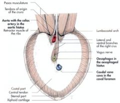
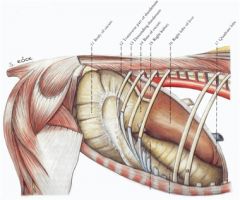
What are the openings of the diaphragm of the horse? What passes through them?
|
Aortic hiatus – between the two crura
Aorta (9) Right azygous v. (10) Thoracic duct (11) Esophageal hiatus – perforates the right crus near its junction with the central tendon Esophagus (14) Dorsal and ventral vagal trunks (12) Esophageal vessels Caval foramen – courses through central tendon toward the right side Caudal vena cava (13) |
|
|
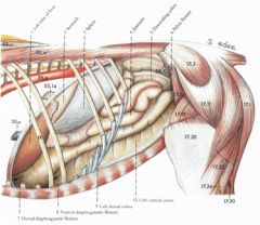
What courses through the diaphragm in the ruminant?
|
Aortic hiatus
Aorta Left azygous v. (it's the right azygous in the horse) Thoracic duct Esophageal hiatus Esophagus Dorsal and ventral vagal trunks Esophageal vessels Caval foramen Caudal vena cava |
|
|
What are the two splenic ligaments?
Which one can cause colic? |
Gastrosplenic ligament
Renosplenic (nephrosplenic) ligament Intestines can get looped over the renosplenic ligament and cause colic. |
|
|
Which liver lobe will you primarily see on the left side of the horse?
What about on the right side? |
Left side: The left lateral lobe.
Right side: Liver – left medial, quadrate, right and caudate lobes |
|
|
What is the analogy to the fundus of the stomach in the horse?
|
The saccus cecus
|
|
|
If you find a smooth muscle band in intestine where do you know you are?
|
The large intestine - they are haustra.
|
|
|
If you're auscultating the right paralumbar fossa of the horse what are you listening to?
|
The base of the cecum.
|
|

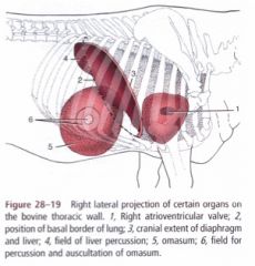
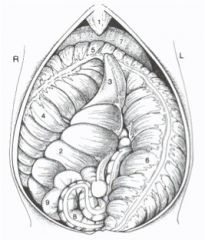
ID 14, 15, 13, 16 in this caudal view of the ruminant.
Where do these structures course between? |
Greater omentum
Superficial leaf (14) Courses from the dorsal body wall on the right side to the left longitudinal groove of the rumen Deep leaf (15) Courses from the dorsal body wall on right side to the right longitudinal groove of the rumen Forms the supraomental recess (13) Omental bursa (16) Space located between the superficial and deep leaves of the greater omentum |
|
|
What does the deep leaf of the greater omentum form a sling for? What recess does it form?
|
Sling for intestines
Forms the supraomental recess. |
|
|
Which species has a thicker greater omentum - small ruminants or ox?
|
Ox
|
|

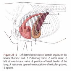
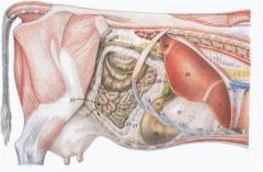
Which of these structures is the reticulum? How do you know?
|
2
It sits next to the diaphragm on the other side of the pericardium. |
|
|
What lobes of the liver can you see on the right side of the ruminant abdomen?
|
Liver – right, quadrate, left, caudate lobes
|
|
|
Is the gall bladder of the ruminant visible on the right or left side?
|
Right
|
|
|
Is the abomasum of the ruminant visible on the right or left side?
|
Right
|
|
|
Is the omasum of the ruminant visible on the right or left side?
|
Right - covered by lesser omentum.
|
|
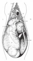
Name!
|

Esophagus (5)
Dorsal sac of rumen (11) Reticulum (6) Omasum (9) Abomasum (10) Descending duodenum (12) Intestinal mass (13) |
|
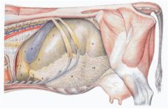
What organs project on the right thoracic wall of the ruminant?
|
What organs project on the left thoracic wall of the ruminant?
|
|
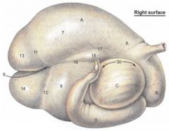
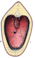
ID this stuff
|

Rumen (A)
Longitudinal groove separates the dorsal (7) and ventral (9) sacs Reticulum (B) Omasum (C) Abomasum (D) |
|
|
Why can't we see things on the right side of the ruminant?
|
The greater omentum covers it all
|
|

What are 3, 4, and 6?
|
3 – apex of cecum
4 - right ventral colon 6 – left ventral colon then transverse colon then descending colon |
|
|
What will you see on the right side equine abdominal topography?
|
Right kidney
Liver – left medial, quadrate, right and caudate lobes Jejunum Cecum Base Body Right ventral colon (part of ascending colon) Right dorsal colon (part of ascending colon) |
|
|
What will you see on the left side of equine abdominal topography?
|
Spleen
Gastrosplenic ligament Left kidney Renosplenic (nephrosplenic) ligament Liver – left lateral lobe Stomach – saccus cecus and body Greater omentum Jejunum Descending colon Left ventral colon (part of ascending colon) Left dorsal colon (part of ascending colon) |
|
|
What will you see on the left side of ruminant abdominal topography?
|

Spleen (4)
Rumen Dorsal sac (7) Ventral sac is covered by greater omentum Left longitudinal groove Caudodorsal blind sac (8) Left dorsal coronary groove Greater omentum (9) Reticulum (2) |
|
|
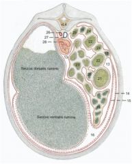
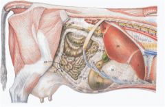
What will you see on the right side of ruminant abdominal topography?
|
Liver – right, quadrate, left, caudate lobes
Gall bladder (27) Right kidney (14) Greater omentum (20,21) Abomasum (29) -Pyloric region (28) Duodenum -Cranial part (26) -Cranial duodenal flexure -Descending duodenum (13) Omasum (30) (covered by lesser omentum) |