Reading...
![]()
Play button
![]()
Play button
![]()
Use LEFT and RIGHT arrow keys to navigate between flashcards;
Use UP and DOWN arrow keys to flip the card;
H to show hint;
A reads text to speech;
35 Cards in this Set
- Front
- Back
|
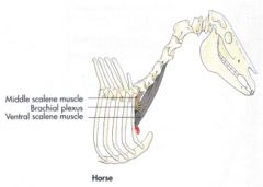
What are the OIAI's of scalenus medius m. in the horse?
|
Scalenus medius m. (both the ventral and middle parts in the picture)
Separated into two parts Cervical roots of the brachial plexus emerge between two parts O: cranial border and lateral surface of the first rib I: dorsal part – transverse process of C7 vertebra I: ventral part – transverse processes of C4-C6 vertebrae Action: flex the neck, bend the neck laterally, draw first rib cranially to assist with inspiration Innervation: ventral brs. of cervical spinal nn. |
|
|
What are the OIAI of the scalenus m. of the ruminant? What are its parts?
|
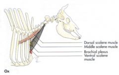
Scalenus dorsalis m. - absent in sheep
O: transverse processes of C4-C6 vertebrae I: 4th rib in ox, 2nd rib in goat Scalenus ventralis m. O: transverse processes of C3-C7 vertebrae I: first rib Action: flex the neck, bend the neck laterally, draw first rib cranially to assist with inspiration Innervation: ventral branches of cervical spinal nn. |
|
|
What are the OIAI of the rectus thoracis m. of the large animal?
|

O: lateral surface of the first rib, ventral to scalenus
I: Horse – cartilage of the 4th rib (may extend to the 5th rib and sternum), aponeurosis joins rectus abdominis I: Ruminant – cartilages of 3rd and 4th or 5th ribs and adjacent parts of sternum Action: draw cartilages and ribs cranially and laterally to assist in inspiration Innervation: intercostal nn. |
|
|
What are the OIAI of serratus dorsalis cranialis m.?
|

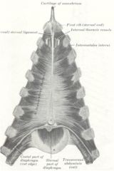
O: thoracolumbar fascia (and dorsoscapular ligament in the horse)
I: lateral surfaces of 5th/6th to 11th/12th ribs in the horse; 6th to 9th ribs in the ruminant Action: draw the ribs cranially and laterally assisting in inspiration Innervation: thoracic spinal nn. |
|
|
What is the OIAI of serratus dorsalis caudalis m.?
|

O: thoracolumbar fascia
I: lateral surfaces of the last 7 or 8 ribs in the horse; last 3-4 ribs in the ruminant Action: draw the ribs caudally assisting with expiration Innervation: thoracic spinal nn. |
|
|
What are the OIAI of the external intercostal mm. of the large animal?
|

O: caudal borders of the ribs
I: cranial borders and lateral surfaces of succeeding ribs Action: draw the ribs cranially to assist with inspiration Innervation: intercostal nn. |
|
|
What are the OIAI of the internal intercostal mm. of the large animal?
|

O: cranial border of the ribs and their cartilages
I: caudal borders of the preceding ribs and cartilages Action: draw ribs caudally to assist with expiration |
|
|
What is the action of the intercostal muscles when working together?
|
Act together to narrow the intercostal spaces and to prevent the thoracic wall from being pushed out or pulled in during respiration
|
|
|
What are the origins of the external abdominal oblique m of the large animals?
|
O: Horse – lateral surfaces of 4th-18th ribs and the fascia over the external intercostal mm.; thoracolumbar fascia
O: Ruminant – caudal border and lateral surface of 6th-13th ribs and the fascia over the external intercostal mm. |
|
|
What is the OIAI of transversus thoracis m.?
|
Located on the thoracic surface of the sternum and the cartilages of the sternal ribs
O: sternum I: costal cartilages and ribs 2-8 Action: draw the ribs and their cartilages medially and caudally, assisting with expiration Innervation: intercostal nn. |
|
|
What are the three epaxial muscles atop the thoracic cavity?
|
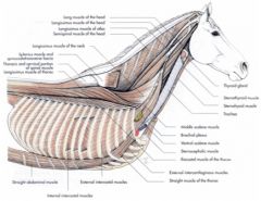
Iliocostalis thoracis m.
Longissimus thoracis m. Transversospinalis muscle system |
|
|
How many ribs do horses and ruminants have?
|
18 ribs in the horse
13 ribs in the ruminant |
|
|
What is the ligament coursing between the heads of the ribs?
|
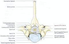
Intercapital ligaments
|
|
|
What are the rib landmarks of the caudal border of the scapula, the supraglenoid tubercle, and the olecranon in the horse and ox?
|

Caudal border of the scapula:
Horse – 7th rib; Ox – 6th rib Supraglenoid tubercle: Horse – just cranial to the 1st rib Olecranon: Horse – 5th rib or 5th IC space ventrally Ox – 5th IC space ventrally |
|
|
How far up does the diaphragm extend into the thoracic cavity of the horse and ox?
|
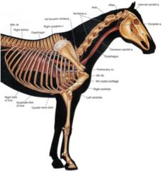
Horse – diaphragm extends cranially to the 6th intercostal space or 6th rib
Ox – diaphragm extends cranially to the 6th rib |
|
|
What are the boundaries of the thoracic cavity of the horse and ox?
|
Cranial boundary – thoracic inlet
Caudal boundary – diaphragm Dorsal boundary – longus colli and thoracic vertebrae Ventral boundary – sternum and transversus thoracis Lateral boundary – ribs and internal intercostal muscles |
|
|
What separates the pleural cavity in the horse and ox? In which species is the separation thicker?
|
Pleural cavities
Only contains a scant amount of serous fluid Separated by the mediastinum Mediastinum is not complete in the horse; delicate Thicker in the ruminant |
|
|
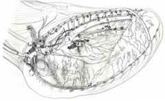
What is in the mediastinum?
|
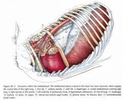
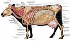
Heart, trachea, esophagus, aorta, lymph vessels, caudal and cranial vena cava, vagus n., phrenic n. (horse shown)
|
|
|
What is the cranial extent of the pleural cavity? What side is it larger on?
|
Cupula Pleura - Cranial extent of the pleural cavity – extends cranial to first rib. It's part of the pleura not in the thoracic cavity.
Larger on the right side |
|
|
What is the line of pleural reflection?
Where is this found in the horse and ox? Why is this must know information? |
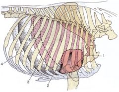
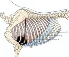
Where the costal pleura is continuous with the diaphragmatic pleura
Horse – from the 8th and 9th costal cartilages dorsocaudally in a gentle curve to the middle of the cranial border of the last rib to the vertebral end of the 17th intercostal space Ox – (indicated as 4 on the picture) from the 8th costochondral junction to the middle of the 11th rib to the 12th rib dorsally Clinically relevant because you'll need to know where to access the thoracic cavity and hit lung, not hit lung, hit abdomen or thorax. MUST KNOW |
|
|
What are the pleural recesses of the large animals?
|
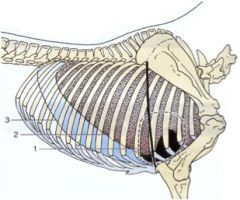
Costomediastinal recess - The blue swath in the picture of the horse
Space ventral to the lungs Costodiaphragmatic recess Space caudal to the basal border of the lungs |
|
|
What are the auscultation borders of the horse and ox?
|
horse: Caudal angle of the scapula, point of the elbow, upper end of the 16th rib
ox: Triceps brachii m., muscles of the back, line from olecranon to the 11th rib dorsally Slightly larger area on right side |
|
|
What is the boundary to perform a pericardiocentesis in the ox? Why are you more likely to do this in an ox then horse?
|
5th IC space, dorsal to costochondral junction, left side
Ruminants are apt to eat something that might puncture their pericardium or heart. |
|
|
What are the boundaries of a thoracocentesis of a horse and ruminant?
|

Horse
Lower part of the 7th IC space, ventral to the margin of the lung Avoid the superficial thoracic vein (spur vein) Ruminant 6th or 7th IC space, dorsal to the costochondral junction |
|
|
T/F The right and left lungs of the equine are equal in size.
|
True
|
|
|
T/F Equine lungs lobes can be seen externally.
|
False
No evidence of external lobation except accessory lobe of right lung |
|
|
What are the basal borders of the equine lung?
What separates the lungs from the line of pleural reflection? |
(line 2 in the picture)
Dorsal region of the 16th rib Middle of the 11th rib Costochondral junction of the 6th rib Separated from the line of pleural reflection by approximately 5 cm dorsally and ventrally, 15 cm in the middle |
|
|
What are the lobes of the right and left equine lung?
Where are the cardiac notches? |
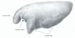
Right:
Cranial, caudal, accessory lobes Cardiac notch 3rd to 4th IC spaces Left (shown): Cranial and caudal lobes Deep cardiac notch 3rd to 6th IC spaces Pericardium contacts the thoracic wall at the 3rd and 6th ribs |
|
|
Are the bovine lungs lobulated? Define their tissue septa.
What defines their basal border? |
Pronounced lobation
Lobulated - thick connective tissue septa Basal border 6th costochondral junction to 11th rib dorsally Straight line |
|
|
Which lung is larger in the bovine? What lobes does it have?
|
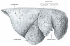
Right lung larger than left lung
Right lobe - Cranial and caudal parts Cardiac notch – 3rd to 4th IC space Middle lobe Caudal lobe Accessory lobe |
|
|
Where is the cardiac notch of the right bovine lung?
The left bovine lung? |
Right Cardiac notch – 3rd to 4th IC space
Left Cardiac notch – 3rd IC space to 5th rib |
|
|
What is unique about the air passage to the right cranial lung lobe of the ruminant?
What is the clinical signifigance of this? |
Tracheal bronchus courses to the right cranial lung lobe
Present in ruminants Want to make sure that the tracheal tube ends before the tracheal bronchus or else the right cranial lung lobe won't get oxygenated. |
|
|
What are the lobes of the left bovine lung?
|
Cranial (with Cranial and caudal parts) and caudal lobe
|
|
|
What is the pathway of lymph drainage from the lungs in the horse?
|
Lymph from the lungs goes to the pulmonary lymph nodes then to the tracheobronchial lymph nodes then to the mediastinal lymph nodes
|
|
|
What are the four lymphocenters of the thoracic cavity of the horse and ox? Which are more developed in the ox?
|
Dorsal thoracic lymphocenter
Intercostal lnn. (4) Thoracic aortic lnn. (5) Ventral thoracic lymphocenter Cranial sternal lnn. (1) Caudal sternal lnn. (1’) Mediastinal lymphocenter Mediastinal lnn. – cranial (2), middle, caudal (6) (caudal mediastinal ln are well-developed in the ox) Nuchal ln. (3) (technically in the neck region) Bronchial lymphocenter Tracheobronchial lnn. – right, left (8), middle (7) Pulmonary lnn. |