![]()
![]()
![]()
Use LEFT and RIGHT arrow keys to navigate between flashcards;
Use UP and DOWN arrow keys to flip the card;
H to show hint;
A reads text to speech;
17 Cards in this Set
- Front
- Back
- 3rd side (hint)
|
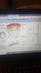
Layers of the retina (in order) |
1.Retinal Pigment Epithelium
2.Photoreceptor layer
3.Outer limiting membrane
4.Outer nuclear layer
5.Outer plexiform layer
6.Inner nuclear layer Horizontal cell Bipolar cell Amacrine cell
7.Inner plexiform layer
8.Ganglion cell layer
9.Nerve fiber layer
10.Inner limiting membrane
Müller cell (throughout the layers) |
|
|
|
Optic disc |
1.5mm
No retinal neurons or RPE
(Blind spot) |
|
|
|
Parafovea and perifovea |
Parafovea -rods and cones -inner and outer nuclear layers 3mm
Perifovea -all retinal layers -thickest part 5mm
|

|
|
|
Macula |
5.5mm Overlaps the fovea, parafovea and perifovea. Contains yellow pigment 'xanthophyl 0.35'
|
|
|
|
Fovea and Foveola |
Fovea 1-2mm Is a depression
Foveola 0.35mm (optimal acuity) Only long-slender cone outer segments |
|
|
|
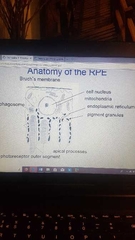
Anatomy of RPE |
-Bruch's membrane -cell nucleus and mitochondria -endoplasmic reticulum -phagosome -pigment granules -photoreceptor outer segments -apical processes |
|
|
|
Functions of RPE |
-light absorption (melanin pigment granules absorb stray light reducing light scatter and improving resolution)
-Epithelial transport (active import from choriocapillaris: glucose, vitamin A and diffusion:H2O and O2. Active export from choriocapillaris: waste products and diffusion: H2O, CO2 ans chloride).
-Active potassium transport (ion levels must be stable for photoreceptors to function. Rapid ion compensation mechanisms in outer retina is done by RPE and müller cells. -Vitamin A metabolism (visual cycle) (retinal is a molecule made by vitamin A. Photons of light are absorbed by transformation of retinal in photoreceptor membranes. Retinal transformed in the rods is restored to its active state by enzymes in RPE.)
-Phagocytes (photoreceptors are produced continuously and shed) rods shed 1-2 hrs after dawn. Cones shed 3-5 hrs after dark. -Hormone production and secretion RPE secretes factors and signalling molecules for regulation of metabolism and reactions to pathology and injury.
|
|
|
|
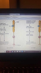
Anatomy of rods, cones and foveola |
Rods 110-130 million per retina Long, slim cells (2 micron diameter) Cones 5-7 millions per retina Wider cells (6 micrkn diameter) Foveola Slim densely packed cone outer segments (1.5 micron diameter) |
|
|
|
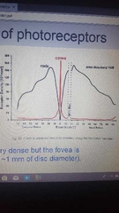
What is the distribution of photoreceptors (cones and rods) like? |
|
|
|
|
What happens when pigment catches a photon |
->decrease [cG] closes cation channels, ->reducing depolarising imward current ->hyperpolarising the cell ->reducing amount of transmitter released |
|
|
|
What is the neural layer made up of |
Photoreceptor layer Outer synaptic layer Bipolar layer (horizontal, bipolar, amacrine cell) Inner synaptic layer Ganglion layer |
|
|
|
Rods and cones |
Both contain photopigments necessary for the absorption of light (that lead to beginning of production of receptor potential). Only rods contain rhodopsin Cones contain 3 different photopigments, one for each type of cone (red, blue and green light sensitive) Photopigments respond to light in a cyclical process |
|
|
|
Phototransduction |
Light adaptation occurs when one moves from dark to light surroundings. (Occurs in seconds)
Dark adaptation occurs when one moves from lit up area to dark one (takes minutes to complete)
This difference is related to rates of bleaching and regeneration of photopigments in rods and cones.
Light causes rod photoreceptors release of excitatory neurotransmitter 'glutamate' to decrease. |
|
|
|
Phototransduction descip. |
In darkness, rod photoreceptors release glutamate. This inhibits bipolar cells from transmitting signals to ganglion cells. Which provide output from retina to the brain.
In light rod photoreceptors are bleached amd hyperpolarise. Glutamate production stops and a reversal occurs on some bipolar cells that are excited into sending signals to ganglion cells which provide output from the retina to brain. Light is the ligand that triggers activation of the enzyme. |
|
|
|
Rod vs cones sensitivity |
-Rods hyperpolarise to very dim light and saturate quickly -cones less sensitive by 2 log units and saturate slowly |
|
|
|
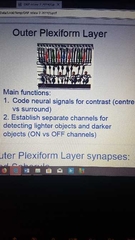
Outer plexidorm layer |
|
|
|
|
Photocurrent from rod and cones |
Rods -slow to respond -very sensitive and adapt only over a small range -are single photons detectors -great amplificatiom takes time to develop and are slow
Cones -respond quickly, biphasically -insensitive and adapt over a very large range of light intensities.
|
|