![]()
![]()
![]()
Use LEFT and RIGHT arrow keys to navigate between flashcards;
Use UP and DOWN arrow keys to flip the card;
H to show hint;
A reads text to speech;
33 Cards in this Set
- Front
- Back
|
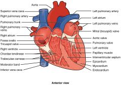
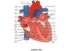
1. Left pulmonary artery 2. Left atrium 3. Left pulmonary veins 4. Mitral (bicuspid) valve 5. Aortic valve 6. Pulmonary valve 7. Left ventricle 8. Papillary muscle 9. Interventricular septum 10. Epicardium 11. Myocardium 12. Endocardium 13. Aorta 14. Superior vena cava 15. Right pulmonary artery 16. Pulmonary trunk 17. Right pulmonary veins 18. Right atrium 19. Foss ovalis 20. Tricuspid valve 21. Right ventricle 22. Chordae tendineae 23. Trabeculae carnea 24. Moderator band 25. Inferior vena cava |
|
|
What do valves in the heart ensure? |
Unidirectional blood flow. |
|
|
What valves prevent backflow of blood? |
Tricuspid valve and pulmonary semilunar valve. |
|
|
What is the role of the chordae tendineae? |
Chordae tendineae prevent AV valve prolapse during ventricular contraction. |
|
|
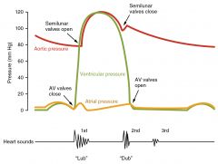
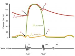
1. Semilunar valves open 2. Semilunar valves close 3. Atrioventricular valves open 4. Atrioventricular valves close 5. Aortic pressure 6. Ventricular pressure 7. Atrial pressure |
|
|
What is the pulmonary circuit? |
The system which runs from the and to the heart from the lungs |
|
|
What is the systemic circuit? |
Heart to and from the tissues in the body. |
|
|
What are the major layers of cardiac muscle? |
1. Endocardium 2. Myocardium 3. Pericardium |
|
|
What are the functions of the pericardium? |
1. Sack for the heart 2. Fixes heart in place 3. Limits motion 4. Protects against infection 5. Lubricates the heart 6. Prevents excess dilation during volume overload |
|
|
Why does the left ventricle have the thickest portion of cardiac muscle of the 4 chambers? |
Left ventricle has thickest in order to generate huge pressure to pump blood through the rest of the body. |
|
|
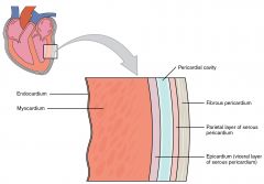
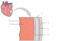
1. Pericardial cavity 2. Fibrous pericardium 3. Parietal layer of serous pericardium 4. Epicardium (Viceral layer of serous pericardium) 5. Myocardium 6. Endocardium |
|
|
What are intercalated disks? |
Junctions between myocardial units. |
|
|
What are intercalated disks made up of? |
Desmosomes and gap junctions. |
|
|
What is the role of desmosomes in the intercalated disk? |
Mechanically hold cardiomyoctes together. |
|
|
What is the role of gap junctions in the intercalated disk? |
Allow passage of ions for electrical travel. |
|
|
1. What are gap junctions made up of? 2. How does the composition of gap junctions vary throughout the heart? |
1. Made up of connexins 2. Vary based on location. Connexin 43 = Ventricular Gap Junction Connexin 40 = Atrial Gap junction |
|
|

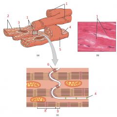
1. Intercalated discs 2. Mitochondria 3. Nucleus 4. Gap Junctions 5. Cardiac Muscle Fibre 6. Desmosome 8. A band 9. I band |
|
|
What is the term given to muscle which can initate its own contraction? |
Myogenic |
|
|
What is the term given to the regular contraction of the heart muscle? |
Autorhythmicity |
|
|
What is autorhythmicity coordinated by? |
Autorhythmic cells. |
|
|
What are the types of autorhythmic cell? |
1. Pacemaker cells 2. Conduction fibres |
|
|
What are the function of pacemaker cells? |
i) Initiate AP ii) establish rhythm iii) Cells of SA and AV node |
|
|
What are the function of conduction fibres? |
i) transmit AP through the heart ii) Cause fast transmission: 4m/second vs 0.5 m/sec for ordinary fibres |
|
|
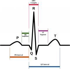
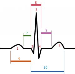
1. P wave 2. Q 3. R 4. S 5 T Wave 6. PR interval 7. PR segment 8. QRS complex 9. ST segment 10. QT interval |
|
|
What does the P wave relate to in terms of the heart beat? |
Activation of the Atria |
|
|
What does the QRS complex relate to in terms of the heart beat? |
The activation of the ventricles |
|
|
What is the T wave otherwise known as? |
Recovery wave. |
|
|
What is special about the length of the potential of the cardiac muscle? |
Cardiac muscle has a prolonged plateau phase (200 millisec) (Normal muscle = 5 millisec) |
|
|
Biochemically, how is the plateau phase of the cardiac muscle extended? |
Calcium ion entry into the autorhythmic cells extends the plateau considerably. |
|
|
What ion movement occurs in the phases of the cardiac action potential? |
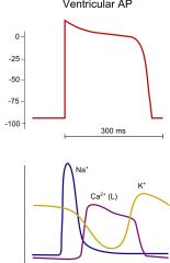
a) Phase 4: Resting phase - K+ channels open b) Phase 0: Depolarisation - K+ channels close and Na+ channels open c) Phase 1 & 2: Repolarisation: K+ channels begin to open - calcium ion channels open d) Phase 3: Calcium ion channels closed. K+ rectifier channels open. e) Phase 4: Return to rest, K+ channels fully open. |
|
|
What is the 'Funny Current'? How does it work? |
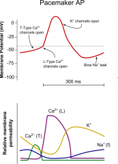
The funny current refers to the slow inward Na+ current which gradually depolarises the membrane of the sinoatrial node membrane at rest. The funny current gradually depolarises the membrane and causes the opening of L-Type and T type voltage gated Calcium ion channels. This generates the 'O' upstroke phase. |
|
|
What does the extended refractory period of the cardiac pacemaker allow for in the heart contraction cycle? |
The refractory period of the cardiac pacemaker lasts almost as long as the entire muscle twitch which allows for specific autonomic timing of the heart rate. |
|
|
How do cytoplasmic calcium levels change over the excitation coupling event? |
1. Excitation events elevate the calcium levels in the cytoplasm i) Calcium enters via voltage gated calcium channels ii) Activation of ER ryanodine receptors releases stored calcium from the sarcoplasmic reticulum. 2. Reductory events - remove calcium ions following excitation. i) Cell surface removal via calcium pumps and the Na+/Ca2+ exchanger (NCX) ii) Sequestration by organelles - Endoplasmic reticulum and mitochondria |