![]()
![]()
![]()
Use LEFT and RIGHT arrow keys to navigate between flashcards;
Use UP and DOWN arrow keys to flip the card;
H to show hint;
A reads text to speech;
37 Cards in this Set
- Front
- Back
|
Neural crest cell formation |
Neural crest cells pinch off from the dorsal neural tube and migrate in a pre set pattern. Grouped based on pinching area and what they form |
|
|
Cranial Neural Crest |
Cartilage, bone, cranial neurons, glia, connective tissues of the face Pharyngial arches --> thymic cells, odontoblasts, bones of the middle ear, jaws |
|
|
Trunk Neural crest |
sensory neurons or melanocytes |
|
|
Vagal and caral neural crest |
parasympathetic ganglia of the gut |
|
|
Cardiac neural crest |
contribute to face structures as well as connective tissue of arteries |
|
|
CNC - intramembranous ossification (alternative) |
Neural crest derived mesenchymal cells condense into nodules where then differentiate to become osteoblasts - bone precursor cells which secrete osteoid matrix that binds Ca Depends on CBFA1 transcription factor (runx2 marker) CBFA1 mutant - cartilage no bone |
|
|
Origin of frontal and parietal bones - mammal |
Neural crest (ectodermal) and mesodermal |
|
|
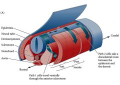
Trunk neural crest cell migration in chick Two pathways |
1. Early: Ventral pathway through anterior, ROSTRAL sclerotome (mesoderm that becomes vertebral cartilage origionate from somites). Become sensory and sympathetic neurons, adrenomedullary cells and schwann cells
2. Late: dorsolateral pathway. Become melanocytes |
|
|
Ventral pathway migration is dependent on components of ECM and chemotactic factors |
ECM modecules in anterior sclerotome permit migration: fibronectin, laminin, collagen, proteoglycan
ECM obstructions: ephrins and semaphorin |
|
|
Dorsal pathway occurs after maturation of dermatomes - mesoderm originating from somites, becoming back dermis |
*timing of somite differentiation regulates spatial and temporal migration of the neural crest cells* Travel through dermis and epidermis to developing hair follicles |
|
|
Cardiac neural crest |
Heart forms in neck region under pharyngeal arch acquiring cells from neural crest Cardiac NCC migrate - bellow otic vesicle, through arches, along aoritc arch arteries, into outflow tract of heart Generate endothelium of aortic arches and septum between aorta and pulmonary artery Pax3 transcription factor |
|
|
Neural plate border: Cranial placodes |
Wnts, BMPs, FGFs and RA induce neural plate border specifier transcription factors (prevent region from becoming neural plate or epidermis) These regulate NCC specifiers (induce neural crest fate) and pre-placodal specifiers |
|
|
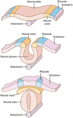
Ectoderm Placode Neural crest Neural groove Notochord |
|
|
Ventral pathway Dorsal pathway Epidermis Neural tube Dermatome Sclerotome Notochord |
|
|
Sensory placodes |
Local transient thickening of the ectoderm in the head and neck Give rise to sensory neurons that form distal portions of ganglia Vision, hearing, balance, taste, smell Proximal portions of ganglia formed from Cranial NCC |
|
|
Lens placodes - reciprocal induction |
Optic vesicle envaginates from diencephalon, contacts ectoderm, induces thickening, forms lens placode, and eventually lens pit |
|
|
Lens pit then induces |
optic vesicle to become optic cup which forms retina optic cup stays connected with diencephalon through optic stalk pit fills in optic cup and closes, detaches from ectoderm and forms lens vesicle |
|
|
Lens vesicle |
Composed of proliferating progenitor lens cells - posterior lens vesicle cells exit mitosis, differentiate -anterior lens vesicle cells remain as monolayer and form anterior lens epithelium |
|
|
Gastrulation and neurulation of the chick embryo |
Notochord extends beneath the neural tube Paraxial mesodem on either side of the neural tube is presomitic mesodem (PSM) somites form from rostral end |
|
|
Somites differentiate into |
Sclerotome Myotome Dermatome |
|
|
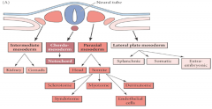
Lateral plate mesoderm splits into |
Splanchinic Somatic Extraembryonic |
|
|
Anterior to the trunk mesoderm is |
Prechordal plate mesoderm |
|
|
Trunk Mesoderm |
1. Chordamesoderm - notochord 2. Paraxial mesoderm - somites form and produce connective tissues of back 3. Intermediate mesoderm - urogenital system, cortical portion of adrenal gland 4. Lateral plate mesoderm - circulatory system, body cavity lining, limbs except for muscle Divisions due to increasing amounts of BMP |
|
|
BMP gradient and mesoderm formation |
High - ventral and lateral - lateral plate mesoderm Lower - intermediate mesoderm Absence - paraxial mesoderm, BMP antagonists such as chordin and nogin |
|
|
intermediate mesoderm chordamesoderm paraxial mesoderm lateral plate mesoderm |
|
|
Paraxial mesoderm becomes the head and somites Sommites become |
Sclerotome - vertebrae, ribs, rib cartilage Myotome - musculature of the back, ribs, limbs Dermatome - dermis of the back Minor components: Syndetome - tendons Endothelial cells - generate vascular cells in dorsal aorta |
|
|
Paraxial mesoderm |
Initially unsegmented, then forms somites for a short time before they develop further # of somites is species specific Somites determine migration path of NCC, spinal nerve axons and form vertebrae, ribs, dorsal dermis, skeletal muscle of back, body and limbs |
|
|
Noggin is responsible for |
Somite formation When inserted into lateral plate mesoderm, somite forming paraxial mesoderm is formed |
|
|
Somitogenesis issues |
1. periodicity 2. fissure formation 3. epithelialization 4. specification 5. differentiation |
|
|
Somite boundries |
quail presumptive boundry cells cause a boundry when transplanted in a chick quail non boundry cells do not *notch signalling pathway helps with boundry formation* quail non boundry cells transplanted but local ectopic expression of notch induced, somite boundry is induced |
|
|
Notch signalling pathway |
Juxtacrine signaling Notch is a receptor on cell membrane, interacts with ligand on adjacent cell, intracellular portion of notch is cleaved and it goes to nucleus, influences gene expression |
|
|
Delta like 3 |
The ligand for notch If mutated it disrupts somite formation Results in rib malformation and aberrant ossigication |
|
|
Hairy 1 |
transcription factor, wave gradient moving caudal to rostral through unsegemented paraxial mesoderm eventually just a thin band at anterior point is left, defines the posterior of the next |
|
|
Clock and wave mechanism form somites |
RA expression follows hensens node regression from A to P, in balance with FGF the trigger Notch the clock stimulates Hairy1 an effector and inhibitor, its own inhibitor Creates wave of gene expression to form anouther somite Simple negative feedback loop |
|
|
Somite separation |
EphrinB2 ligand at the posterior of the anterior somite interacts with EphA4 receptor, tirosine kinase in the unsegmented mesoderm |
|
|
Gap forms in the presomitic mesoderm |
Anterior cells to the gap ungergo epithelialization first become posterior somite Posterior cells of the gap undergo epithelialization later MET - mesenchymal to epithelial transition create mesodermal somites surrounded in epithelium |
|
|
MET in peripheral somatic cells |
Caused by ectodermal signals Cells polarize - sub apical surface inward, basal membrane outside Cells synthesize ECM fibronectin protein and Ncadherin |