![]()
![]()
![]()
Use LEFT and RIGHT arrow keys to navigate between flashcards;
Use UP and DOWN arrow keys to flip the card;
H to show hint;
A reads text to speech;
146 Cards in this Set
- Front
- Back
|
Functions of CSF |
Protects brain & spinal cord from injury (shock absorption) |
|
|
Structures of Blood Brain Barrier (BBB) |
Astrocytes & Pericytes surround blood vessels of the brain & spinal cord, helping to control influx/efflux of chemicals Microglia are specialized immune system cells that patrol the brain & spinal cord who look for & remove invaders, damaged cells, or cancerous cells |
|
|
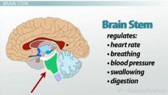
Brain Stem functions |
|
|
|
Brain Stem structures |

|
|
|
What are the functions of the Reticular Activating System (RAS)? |
In human biology, believed to play a role in many important functions, including sleep and waking, behavioral motivation, breathing, and the beating of the heart.
|
|
|
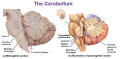
What are the functions of the cerebellum? |
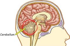
Coordinates/regulates voluntary movements such as posture, balance, coordination, and speech, resulting in smooth and balanced muscular activity Stores learned motor movement |
|
|
What is the function of the thalamus? |
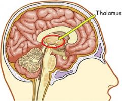
Relays sensory info from sensory receptors to proper areas of the brain where it can be processed (switchboard) |
|
|
What is the function of the hypothalamus? |
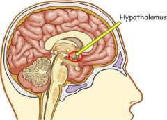
Responsible for motivational behaviors (e.g., hunger, thirst) Maintain constant body temperature Controls pituitary gland, which is the master gland that controls other endocrine glands in the body |
|
|
What is the function of the medulla oblongata? |
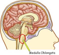
Controls involuntary body functions that sustain life (e.g., breathing, swallowing, heart rate) Helps transfer neural messages from the brain to the spinal cord |
|
|
What are the 5 lobes of the cerebellum? |
|
|
|
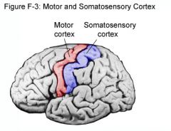
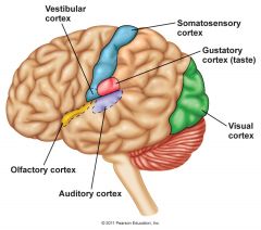
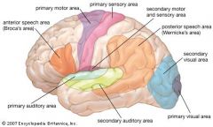
Somatosensory Cortex function & location |

Location: Posterior to the central sulcus, it's made up of right & left lobes which are connected in the middle by the corpus colossum. Function: Receives all sensory input from the body. |
|
|
Primary Motor Cortex function & location |

Location: Anterior to the central sulcus, back of frontal lobe, just about at the top of the head Function: Generate neural impulses that control the execution of movement |
|
|
Pre-frontal Area function & location |
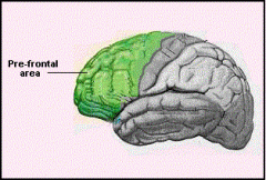
Function: Part of brain that gives human beings much of their intelligence & problem solving ability because its ability to process both current environment & past memories |
|
|
Broca's Area function & location |
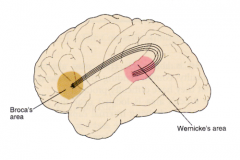
Speech production, language processing |
|
|
Wernicke's Area function & location |

Comprehension of speech |
|
|
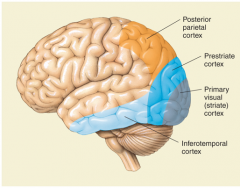
Primary Visual Cortex function & location |
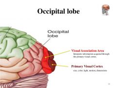
Location: Posterior of Occipital Lobe Function: Photoreception; central processing of visual information |
|
|
Primary auditory area function & location |
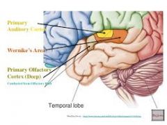
Function: Sound information processed, including: - Frequency - Location - Volume |
|
|
Visual association area function & location |

Location: Directly anterior to the Primary Visual Cortex on the Occipital Lobe Function: Complex processing of visual information |
|
|
Auditory association area function & location |
Function: Complex processing of auditory info; allows you to recognize a particular sound as speech, music, or noise |
|
|
What is a commissural tract? |
Connects LEFT & RIGHT hemispheres of the brain (e.g., corpus collosum) |
|
|
What is an association tract? |
Connect 1 gyrus to another in the SAME HEMISPHERE (e.g., Cingulum) |
|
|
What is a projection tract? |
Connect cerebral cortex to spinal cord and other lower brain structures Run VERTICALLY - convey sensory and motor information |
|
|
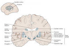
Structures of the basal ganglia (basal nuclei) |

|
|
|
What is the function of the Caudate Nucleus? |
Storing & processing memories Using past to determine future action Language control |
|
|
What is the function of the Globus Pallidus? |
Regulation of voluntary movement |
|
|
What is the function of the Putamen? |
Aids smooth, predictable movement of limbs |
|
|
What is Parkinson's & what are the symptoms? |
Parkinson’s disease affects the nerve cells in the brain that produce dopamine Symptoms: - Muscle rigidity - Tremors - Changes in speech & gait |
|
|
Whydoes L-Dopa improve the symptoms of Parkinson’s Disease? |
Nerve cells use L-Dopa to make dopamine to replenish the brain's dwindling supply |
|
|
Cerebrum |
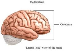
"Seat of intelligence" Reading, writing, speaking |
|
|
Cerebral Cortex |
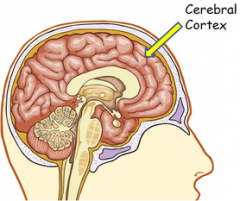
Grey matter on outer seat of cerebellum |
|
|
Gyrus |
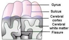
Ridge/fold between 2 clefts on the cerebral surface in the brain |
|
|
Sulcus |

Groove/furrow between folds |
|
|
Longitudinal fissure |
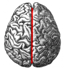
Separates the cerebrum into RIGHT & LEFT halves Contains falx cerebri |
|
|
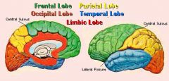
Central sulcus |
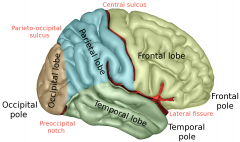
Separates the FRONTAL LOBE & the PARIETAL LOBE |
|
|
Precentral gyrus |
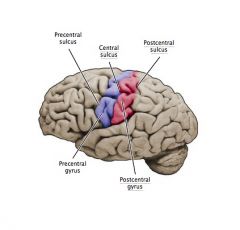
Contains PRIMARY MOTOR area of CEREBRAL CORTEX; Immediately anterior to central sulcus |
|
|
Postcentral gyrus |

Contains PRIMARY SOMATOSENSORY area of CERBRAL CORTEX; Immediately posterior to central sulcus |
|
|
Lateral sulcus |

Separates FRONTAL LOBE from TEMPORAL LOBE |
|
|
Parieto-occipito sulcus |

Separates PARIETAL LOBE from POSTERIOR OCCIPITAL LOBE |
|
|
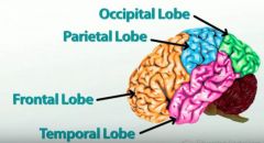
Frontal lobe |

Controls important cognitive skills; Motor association area |
|
|
Prefrontal cortex |
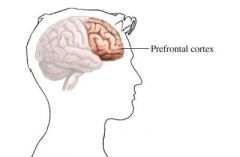
Involves planning & a person's response to complex problems |
|
|
Primary motor cortex |

Plan & execute movement; Precentral gyrus |
|
|
Parietal lobe |
Sensation & perceptions |
|
|
Primary somatosensory area |
Receive nerve impulses for touch, pressure, pain, & temperature |
|
|
Primary gustatory area |
Receive impulses for taste (taste perception) |
|
|
Primary visual area (of occipital lobe) |

Receives visual input |
|
|
Visual association area (of occipital lobe) |
Interprets visual input |
|
|
Temporal lobe
|

Understanding speech; Lies directly deep to temples |
|
|
Primary auditory area |

Receives auditory info; Basic & higher functions of hearing |
|
|
Auditory association area |
Interprets acoustic signals as speech, music, or other sound |
|
|
Primary olfactory area |

Olfactory perception |
|
|
Insula |

"Island;" Integrates ANS info |
|
|
Amygdala |
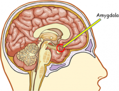
Emotions (response to anger); Survival instincts |
|
|
Hippocampus |
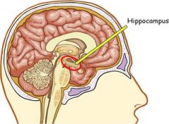
Regulate emotion; Memory & orientation |
|
|
Olfactory bulb |
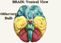
Responsible for sense of smell |
|
|
Basal nuclei |
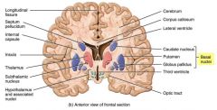
Initiation & termination of movement |
|
|
Claustrum |
Essential in multisensory integration |
|
|
Internal capsule |
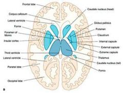
White matter projection fibers; Contains both ascending & descending axons |
|
|
External capsule |

White matter projection fibers; High concentration of motor & sensory projection fibers |
|
|
Extreme capsule |

White matter projection fibers between Broca's and Wernicke's speech areas (Role in language?) |
|
|
Choroid plexus |
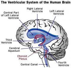
Produces CSF |
|
|
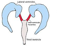
Lateral ventricle |

Contains & circulates CSF |
|
|
Interventricular foramen |
Connects ventricles & allows passage of CSF |
|
|
3rd Ventricle |

Contains & circulates CSF |
|
|
Cerebral aquaduct |
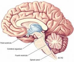
Transfers CSF from 3rd ventricle to 4th ventricle |
|
|
Median aperature |
Drains CSF from 4th Ventricle to Cisterna Magna |
|
|
Lateral aperature |

Drains CSF from 4th Ventricle into Cerebellopontine angle cistern |
|
|
Subarachnoid space |
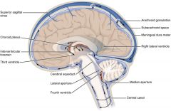
Contains blood vessels (that supply brain) & CSF; Helps cushion brain from injury; Part of BBB |
|
|
Central canal |

CSF-filled space that runs length of spinal cord |
|
|
Arachnoid villi (AKA - Arachnoid granulation) |

Small protrusions in arachnoid mater through dura mater that allows CSF to exit subarachnoid space to blood stream |
|
|
Thalamus |

Involved with motor control; Receives/relay signals to/from cerebral cortex (switchboard); Controlling sleep/wake cycle |
|
|
Intermediate mass |
Connection between right & left portions |
|
|
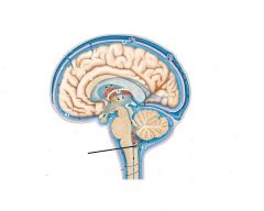
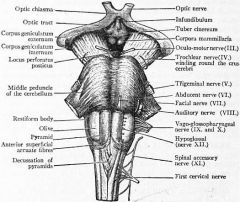
Optic nerve |

Visual pathway |
|
|
Optic chiasma |
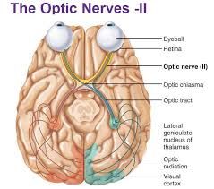
Visual pathway |
|
|
Optic tract |

Visual pathway |
|
|
Anterior commisure |
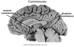
Commissural fibers connecting cerebral hemispheres
|
|
|
Posterior commisure |

Commisural fibers crossing the midline bilateral; Involved in light touch |
|
|
Mammillary body |
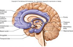
Smell relay, suckling reflex |
|
|
Pineal body (epiphysis) |
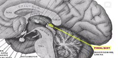
Secretes melatonin;
Regulates endocrine functions; Body clock |
|
|
Pituitary (hypophysis) |
Secretes hormones that control endocrine glands for regulation of all aspects of growth, development, metabolism, & homeostasis |
|
|
Substantia nigra |
Plays role in reward, addiction, & movement via dopamine production |
|
|
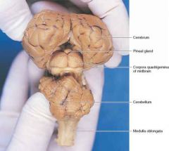
Corpora quadrigemina |
Reflex centers involving vision & hearing |
|
|
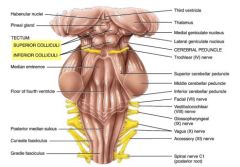
Superior colliculi |
Plays role in helping orient head & eye to all types of sensory stimuli (VISUAL REFLEX) |
|
|
Inferior colliculi |

Principal midbrain nucleus of the auditory pathway (AUDITORY REFLEX) |
|
|
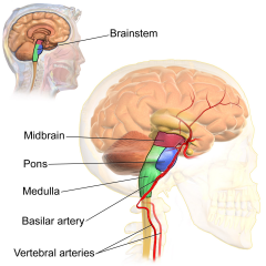
Pons |
Helps relay messages from cortex & cerebellum; Contains breathing centers |
|
|
Middle cerebellar peduncle |

Connects pons to cerebellum; Voluntary movement pathway |
|
|
Cerebellum |
Coordinates & regulates muscular activity |
|
|
Folium |
Folds of the cerebellum |
|
|
Vermis |
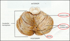
Posterior fossa in the brain that separates Right & Left; Works in posture & locomotion |
|
|
Medula oblongata |

Center for respiration & circulation; Helps regulate digestion, coughing, sneezing, vomiting, & swallowing |
|
|
Olive (olivary bodies) |

Proprioception |
|
|
Decussation of the Pyramids |
Motor tracts of the medulla |
|
|
Falx cerebri |

Extension of dura mater that separates 2 hemispheres of cerebrum |
|
|
Falx cerebelli |

Extension of dura mater that separates the 2 hemispheres of the cerebellum |
|
|
Tentorium cerebelli |

Extension of dura mater that separates cerebrum from cerebellum |
|
|
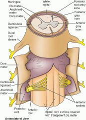
Dura mater |

Most superficial of the 3 meninges composed of dense irregular CT |
|
|
Arachnoid mater |
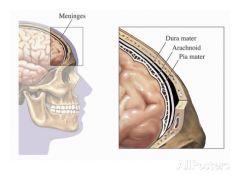
Middle of the 3 meninges; Avascular CT covering |
|
|
Pia mater |

Innermost of 3 meninges; Thin transparent areolar CT layer that adheres to surface of spinal cord & brain |
|
|
Epidural space |
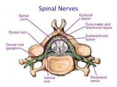
Space between dura mater & wall of vertebral canal |
|
|
Subdural space |
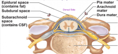
Space between dura mater & arachnoid mater containing interstitial fluid |
|
|
Subarachnoid space |

Space between arachnoid mater which cointains CSF; Shock absorption & suspension system for brain & spinal cord |
|
|
Grey matter |
Unmyelinated axons, mostly cell bodies |
|
|
|
Myelinated axons (tracts)
|
|
|
Anterior median fissure |
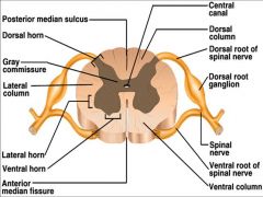
Wide groove |
|
|
Posterior median sulcus |

Narrow furrow |
|
|
Anterior white column |

Somatic motor neurons/axons |
|
|
Posterior white column |
Somatic sensory neurons/axons |
|
|
Lateral white column |

Lots of tracts |
|
|
Anterior grey horn |
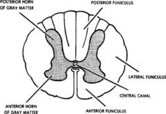
Somatic motor neurons/cell bodies |
|
|
Posterior grey horn |

Somatic sensory neurons/cell bodies (& ANS) |
|
|
Lateral grey horn |
(Only in thoracic & lumbar) ANS motor neurons |
|
|
Grey commissure |

Connects 2 sides of grey matter |
|
|
Central canal |

Contains CSF |
|
|
Epyndymal cells |
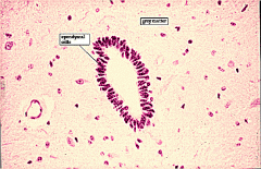
Line central canal; Produce & circulate CSF |
|
|
Anterior horn cells |
Somatic motor neurons |
|
|
Posterior root |
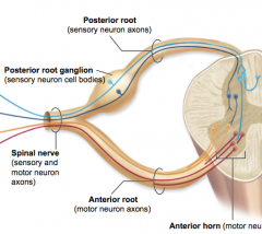
Contains sensory axons |
|
|
Anterior root |

Contains motor axons |
|
|
Posterior root ganglion (DRG) |

Sensory cell bodies |
|
|
Dorsal ramus |
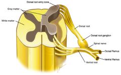
Mixed nerve (motor & sensory) to POSTERIOR
|
|
|
Ventral ramus |

Mixed nerve (motor & sensory) to ANTERIOR |
|
|
Spinal nerve |
Mixed nerve |
|
|
Cervical (brachial) enlargement |

Thickening of spinal cord containing nerves to upper extremities |
|
|
Lumbrosacral (lumbar) enlargement |
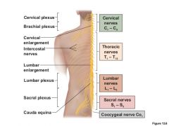
Thickening of spinal cord containing nerves to lower extremities |
|
|
Cauda equina |

Nerves exiting spinal cord resembling horse's tail |
|
|
Filum terminale |

Pia matter extension attaching to coccyx |
|
|
Conus medullaris |
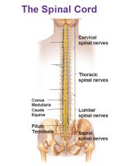
End of spinal cord |
|
|
Nerve of Cervical Plexus (C1-C5) |
Phrenic nerve |
|
|
Trunks of Brachial Plexus (C5-T1) |
Superior, medium, & inferior trunks |
|
|
Cords of Brachial Plexus (C5-T1) |
Posterior, lateral, & medial cords
|
|
|
Lumbar Plexus |
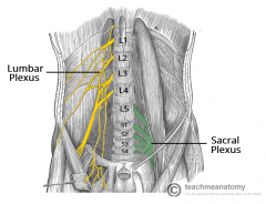
L1-L5 |
|
|
Sacral Plexus |

L4-S4 (e.g., sciatic nerve) |
|
|
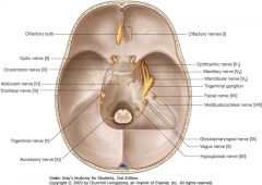
Which cranial nerve exit brain through Telencephalon? |

(I) Olfactory nerve |
|
|
Which cranial nerves exit brain through Diencephalon? |
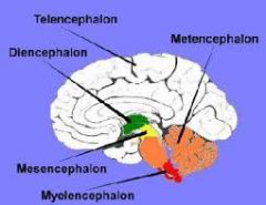
(II) Optic |
|
|
Which cranial nerves exit brain through the Mesensephalon midbrain? |

(III) Occulomotor (IV) Trochlear |
|
|
Which cranial nerves exit brain through the Mesensephalon pons? |

(V) Trigeminal (VI) Abducens (VII) Facial (VIII) Vestibulochoclear |
|
|
Which cranial nerves exit brain through the Mylencephalon medulla? |

(IX) Glossopharyngeal (X) Vagus (XI) Accessory (XII) Hypoglossal |
|
|
What is the skull exit of the olfactory nerve? |
Cribriform plate of ethmoid bone |
|
|
What is the skull exit of the optic nerve? |
Optic foramen |
|
|
Which cranial nerves exit through the Superior Orbial Fissure? |
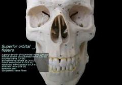
(III) Oculomotor (IV) Trochlear (V) Trigeminal (opthamalic) (VI) Abducens |
|
|
Which skull feature does the Trigeminal maxillary cranial nerve exit out of? |
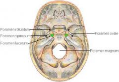
Foramen rotundum |
|
|
Which skull feature does the Trigeminal madibular cranial nerve exit out of? |

Foramen ovale |
|
|
Which skull feature does the Facial cranial nerve exit out of? |
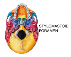
Stylomastoid foramen |
|
|
Which skull feature does the Vestibulochoclear cranial nerve exit out of? |
Internal acoustic meatus |
|
|
Which cranial nerves exit the skull through the Jugular Foramen? |
(IX) Glossopharengeal (X) Vagus (XI) Accessory |
|
|
Which skull feature does the Hypoglossal cranial nerve exit out of? |
Hypoglossal cannal |
|
|
What are the stages of the reflex arc? |
1. Stimulus receptor 2. Sensory neuron 3. Integration center 4. Motor neuron 5. (Reflex) effector |
|
|
Denticulate ligament |
Ligament that keeps spinal cord aligned horizontally |