![]()
![]()
![]()
Use LEFT and RIGHT arrow keys to navigate between flashcards;
Use UP and DOWN arrow keys to flip the card;
H to show hint;
A reads text to speech;
400 Cards in this Set
- Front
- Back
|
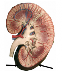
What are the functions of the kidneys? |
--Regulate blood volume & pressure --Regulate pH --Regulate ionic composition --Reabsorption/production of glucose --Excrete wastes --Production/release of hormones (e.g., erythropoietin) |
|
|
What is the renal hilium? |
Indentation in kidney thru which ureter, blood vessels, lymphatic vessels, & nerves emerge from kidney |
|
|
What are the coverings to the kidney? |
--Renul capsule (deep) --Adipose capsule (middle) --Renal fascia (superfiscial) |
|
|
What is the function of the renal capsule? |
--Barrier against trauma --Helps maintain shape |
|
|
What tissue comprises the renal capsule? |
Dense irregular CT |
|
|
What is the function of the adipose capsule? |
--Protect kidney from trauma --Holds kidney in place within abdominal wall |
|
|
What kind of tissue comprises the adipose capsule? |
Adipose tissue |
|
|
What is the function of the renal fascia? |
Anchors kidney to peritoneum |
|
|
What tissue type comprises the renal fascia? |
Dense connective tissue |
|
|
What is the renal cortex? |
Outer region of kidney |
|
|
What is the function of the renal cortex? |
Place of filtrate production (via nephrons) |
|
|
What is the renal medulla? |
Inner region of kidney |
|
|
What is the function of the renal medulla? |
Collection & excretion of urine (via pyramids & columns) |
|
|
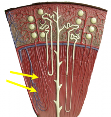
What are the renal pyramids? |
Cone-shaped structures within medulla |
|
|
Where are the renal pyramids located? |
Medulla (of kidney) |
|
|
What structures comprise the renal pyramids? |
Tubules & ducts |
|
|
What are renal papilla? |
Apex of renal pyramids that lead to ducts |
|
|
What are the renal columns? |
Portions of renal cortex that extend between renal pyramids |
|
|
How many renal pyramids does a kidney contain? |
8 - 18 |
|
|
What are the functions of renal columns? |
--Allows cortex to be anchored (via fibrous material) --Contains blood vessels |
|
|
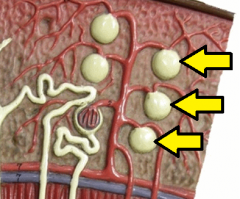
What is the renal cavity? |
Large cavity that drains urine from major calyces to ureter |
|
|
What is a minor calyx? |
Cuplike strcuture that receives urine from papillary ducts of 1 renal papilla |
|
|
How many minor calyces are there per kidney? |
8 - 18 |
|
|
What are major calyces? |
Cuplike strcutures that recieve urine from minor calyces |
|
|
How many major calyces are there per kidney? |
2 - 3 |
|
|
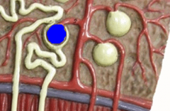
What is the renal sinus? |
Space inside kidney that contains part of renal pelvis calyces, renal blood vessel branches, & nerves |
|
|
How much of cardiac output passes through kidneys? |
33% |
|
|
What is the function of the renal artery? |
Transports oxygen-rich blood into kidney |
|
|
What is the function of the renal vein? |
Transports oxygen-poor blood from the kidneys |
|
|
What is the function of the segmental artery? |
Supply different areas of kidney w/ oxygen-rich blood |
|
|
What is the function of the segmental vein? |
Transport oxygen-poor from different areas of kidney |
|
|
What is the segmental artery/vein? |
Branch of segmental artery/vein that enters parenchyma & passes thru renal column |
|
|
What are the arcuate arteries/veins? |
Arches at base of renal pyramid |
|
|
Where are the interlobular arteries/veins located? |
Between lobules |
|
|
Where are afferent arterioles located? |
In renal cortex (gives rise to glomerulus) |
|
|
What are the functions of efferent arterioles? |
--Place of filtration --Reunite & carry blood out of glomerulus |
|
|
What are the pentubular capillaries? |
Surrounds tubular part of the nephron in the renal cortex (from efferent arterioles) |
|
|
What renal covering is continuous w/ the ureter? |
Renal capsule |
|
|
What are the vasa recta? |
Supplies tubular portion of nephron in renal medulla from efferent arterioles |
|
|
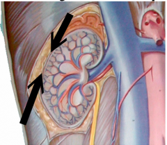
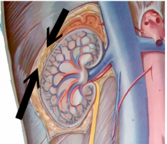
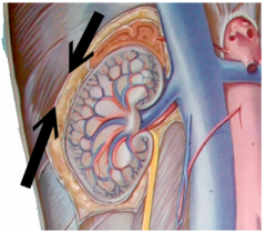
What is the position of the kidneys? |
Retroperitoneal |
|
|
What is the renal corpuscle? |
Part of nephron where blood is filtered |
|
|
What is the glomerulus? |
Component of renal corpuscle capillary network from afferent to efferent |
|
|
What 3 layers comprise the filtration membrane? |
--Fenestrated glomerular endothelial cell --Glomerular basil lamina --Slit membrane (between pedicels) |
|
|
What is the function of fenestrated glomerular endothelial cells? |
Prevents filtration of blood cells, but allows other components of plasma to pass thru |
|
|
What is the function of the glomerular basil lamina? |
Prevents filtration of lg. proteins |
|
|
What is the function of the slit membrane (between pedicels)? |
Prevents filtration of med. proteins
|
|
|
What is the basal lamina (of renal corpuscles)? |
2 fused basement membranes (e.g., b/w red & green screens) of capillary cells & podocytes in filtration membrane |
|
|
What is Bowman's capsule? |
Double walled epithelial cup surrounding glomerulus |
|
|
What layers comprise Bowman's capsule? |
--Visceral layer (proximal to glomerulus) --Parietal layer (distal to glomerulus |
|
|
What comprises the visceral layer of Bowman's capsule? |
Podocytes on top of capillaries |
|
|
What is a podocyte? |
Simple squamous epithelial cells located in visceral layer of Bowman's capsule |
|
|
What kind of tissue are podocytes comprised of? |
Simple squamous epithelial tissue |
|
|
What are pedicels? |
Foot-like processes extended from each podocyte wrapped around glomerular capillaries |
|
|
What are filtration slits? |
Spaces in between pedicels |
|
|
What is the function of filtration slits? |
Prevent med. molecules from exiting |
|
|
What are slit membranes? |
Web-like structure that extends across filtration slits & allows glucose, amino acids, urea, etc. to leave |
|
|
What is the basal lamina (of renal corpuscle)? |
Selective filter that determines which molecules diffusing from underlying CT may enter epithelium |
|
|
What is the function of the filtration membrane? |
Allows free passage of water molecules via podocytes & endothelial cells |
|
|
What is the proximal convoluted tubule? |
Structure immediately following Bowman's capsule |
|
|
What is the function of the proximal convoluted tubule (PCT)? |
Major area of reabsorption: --H2O (66%) --Glucose & amino acids (almost 100%) --Salts |
|
|
What tissue type comprises wall of the PCT? |
Cuboidal epithelial tissue |
|
|
What is the function of the (renal) brush border? |
Increase surface area for reabsorption |
|
|
Approximately how much H2O is reabsorbed in the Loop of Henle? |
15% |
|
|
Approximately how much H2O is reabsorbed in the PCT? |
66% |
|
|
What is obligatory water reabsorption? |
H2O reabsorbed w/ solutes in tubular fluid (because H2O is "obliged" to follow solutes when they're reabsorbed) |
|
|
Where does obligatory water reabsorption occur? |
--PCT --Descending Loop of Henle |
|
|
What is facultative water reabsorption? |
Water absorption that is regulated based on perceived need by ADH
|
|
|
Where does facultative water reabsorption occur? |
Collecting ducts |
|
|
What tissue comprises the descending loop of Henle? |
Simple squamous epithelial tissue |
|
|
What tissue comprises the thin ascending loop of Henle? |
Simple squamous epithelial tissue |
|
|
What tissue comprises the thick ascending loop of Henle? |
Simple cuboidal to low columnar epithelial cells |
|
|
What is the distal convoluted tubule (DCT)? |
Tubule in renal cortex attaching ascending thick LOH to collecting tubules
|
|
|
What is the function of the DCT? |
Some reabsorption based on body's needs |
|
|
What is the function of the collecting duct? |
--Secretion of H+ --Facultative H2O reabsorption |
|
|
What structures for the juxtaglomerular apparatus? |
--Macula densa --Juxtaglomerular cells (in tunica media of afferent arteriole) --Extraglomerular mesangial cells |
|
|
What is the macula densa? |
Where cells of ascending limb of LOH contact afferent arterioles |
|
|
What is the function of mesangieal cells? |
Regulate blood flow of the glomerular capillaries via contractile activity
|
|
|
Where are juxtaglomerular cells located? |
In tunica media of afferent arteriole near ascending LOH |
|
|
What is the function of the ureter(s)? |
Transport urine from renal pelvis to urinary bladder |
|
|
What is the urinary bladder? |
Hollow, distendable organ in pelvic cavity |
|
|
What is the function of the urinary bladder? |
Stores urine |
|
|
What is the orifice of ureter? |
Opening of ureter to urinary bladder |
|
|
What is the trigone? |
Sm. triangular area in floor of urinary bladder containing orifices of ureters |
|
|
What is the detrusor muscle? |
Muscularis of the urinary bladder |
|
|
What are rugae? |
Folds in the mucosa |
|
|
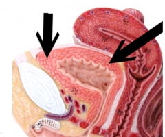
What is the urethra? |
--Terminal portion of urinary system --Passageway for discharging urine from body |
|
|
What is the opening into the urethra? |
Internal urethral orifice |
|
|
Is the internal urethral sphincter voluntary or involuntary? |
Involuntary |
|
|
What is the circular band of muscles around the opening to the urethra? |
Internal urethral sphincter |
|
|
What is the opening inferior to the internal urethral sphincter? |
External urethral orifice |
|
|
What is the circular band of muscles inferior to the internal urethra sphincter? |
External urethra sphincter |
|
|
Is the external urethral sphincter voluntary or involuntary? |
Voluntary (skeletal muscle) |
|
|
What is the prostatic urethra? |
The portion of the urethra that passes thru the prostate (Males only) |
|
|
What is the membranous urethra? |
--Shortest portion of urethra --Portion that passes thru deep muscles of perineum |
|
|
What is the cavernous urethra? |
Longest portion of urethra |
|
|
Trace a drop of blood through the kidney to the peritubular capillaries |
Renal artery --> Segmental arteries --> Interlobar arteries --> Arcuate arteries --> Interlobular arteries --> Afferent arterioles --> Glomerular capillaries --> Efferent arterioles --> Peritubular capillaries |
|
|
Trace a drop of blood through the kidney from the peritubular capillaries |
Peritubular capillaries --> Interlobular veins --> Arcuate veins --> Interlobar veins --> Renal vein |
|
|
How are glomerular capillaries unique among body capillaries? |
They're positioned between 2 arterioles rather than between an arteriole & a venule |
|
|
What is dialysis? |
Filtration |
|
|
What is the space between the 2 layers of the glomerular capsule called? |
Bowman's space -or- Capsular space |
|
|
What is the function of the epithelium of the renal corpuscle? |
Dialysis membrane formed by fenestrations in the epithelium |
|
|
What is the advantage of nephrons with long LOH? |
Enable kidneys to create concentration gradient in renal medulla to excrete very dilute/concentrated urine |
|
|
What are the basic functions performed by nephrons & collecting ducts? |
--Glomerular filtration --Tubular reabsorption --Tubular secretion |
|
|
What is glomerular filtration? |
Formation of a protein-free filtrate (ultrafiltrate) of plasma across glomerular membrane |
|
|
What is the functional portion of the kidney? |
Parenchyma |
|
|
What features does the renal parenchyma include? |
--Renal cortex --Renal pyramid |
|
|
What structures are contained in a kidney lobe? |
--Pyramid --1/2 of adjacent columns --Cortex |
|
|
What happens to plasma & its elements that escape filtration? |
--Exits renal corpuscle via efferent arteriole --Perfuses tubular capillary bed --Collected in renal venous system |
|
|
Which renal cells contain receptors for ADH & where are they located? |
Principal cells: --DCT --Collecting duct |
|
|
What is the function of renal intercalated cells? |
Regulate blood pH homeostasis |
|
|
How do ureters move urine into the bladder? |
Peristalsis |
|
|
What is the function of the juxtaglomerular apparatus? |
Hormonal regulation of... --BP --Renal blood flow --GFR |
|
|
What is the function of juxtaglomerular cells? |
Secrete renin to help increase BP |
|
|
What is the function of a macula densa cell? |
Monitor change in Na+ concentrations in DCT to regulate GFR |
|
|
Which renal cells possibly excrete erythropoietin? |
Extraglomerular mesangial cells |
|
|
What are the 2 countercurrent mechanisms of the kidney? |
--Countercurrent multiplication --Countercurrent exchange |
|
|
What is the function of RAAS? |
Regulation of... --Systemic blood flow --Renal blood flow --GFR |
|
|
What is renin? |
Enzyme released by juxtaglomerular cells in response to low BP |
|
|
What is the function of renin? |
Transformation of angiotensinogen to angiotensin I |
|
|
What is the function of aldosterone? |
--Increase reabsorption of H2O from DCT & collecting ducts --Secretion of K+ into urine |
|
|
What is the function of ADH? |
increase BP |
|
|
What signals the release of ADH? |
Presence of Angiotensin II |
|
|
What structures make up the urinary system? |
--Kidneys --Ureters --Bladder --Urethra |
|
|
How much of the cardiac output (CO) do the kidneysreceive?
|
25 - 33% |
|
|
What are some specific functions of the kidneys? |
--Regulate blood volume, BP, & pH --Regulating ion composition/concentration --Reabsorption/production of glucose --Excreting wastes --Production/release of hormones |
|
|
What is calcitriol? |
Active form of Vitamin D that leads to reabsorption of Ca2+ from ultrafiltrate passing thru renal tubules
|
|
|
What is the function of EPO? |
(Erythropoietin) Promotes formation of RBCs by blood marrow |
|
|
What is the functional unit of the kidney? |
Nephron
|
|
|
What are the 3 layers of tissue surrounding each kidney? |
--Renal capsule (DICT) --Adipose capsule --Renal fascia (DCT) |
|
|
What is the layer of the kidney that is continuous with ureters? |
Renal capsule |
|
|
What is the function of the adipose capsule? |
--Protect kidney from outside forces --Hold kidney in place |
|
|
What is the function of the renal fascia? |
Anchors kidney to peritoneum |
|
|
What is the function of the renal cortex? |
Filtration to form urine (via nephrons) |
|
|
What is the function of the renal medulla? |
Collection & excretion of urine (via pyramids & columns) |
|
|
What are the renal subdivisions that contain the kidney's secreting apparatus & tubules? |
Pyramids |
|
|
How many pyramids does each kidney have? |
8 - 18 |
|
|
What is the fibrous material of the kidney that allows the cortex to be anchored & contains renal blood vessels? |
(Renal) columns |
|
|
What structures make up a nephron? |
--Renal corpuscle --Renal tubules |
|
|
What are the structures that make up a renal corpuscle? |
--Glomerulus --Bowman's capsule |
|
|
What are the 3 types of renal tubules? |
--PCT --LOH --DCT |
|
|
How many nephrons per kidney? |
1 million |
|
|
What are the 2 types of nephrons? |
--Corical nephrons (80 - 85%) --Juxtamedulary nephrons (15 - 20%) |
|
|
Where are cortical nephrons located? How deep does the LOH extend? |
Located in outer portion of cortex w/ short LOH that penetrates only sm. way into medulla
|
|
|
Where do cortical nephrons receive their blood supply from? |
Peritubular capillaries (arising from efferent arterioles) |
|
|
Where are juxtamedullary nephrons located? How deep does the LOH extend? |
Deep in cortex (next to medulla) w/ long LOH that extend into deepest region of medulla |
|
|
Where do juxtamedullary nephrons receive their blood supply from? |
Vasa recta arrising from peritubular capillaries before becoming peritubular venules |
|
|
What is a typical GFR for males? For females? |
Males: 125 mL/min Females: 105 mL/min |
|
|
What are the 2 types of reabsorption? |
--Paracellular reabsorption --Transcellular reabsorption |
|
|
What is paracellular reabsorption? |
Passive process occurring between adjacent tubule cells |
|
|
What is transcellular reabsorption? |
Movement thru an individual cell |
|
|
What is the formula for Net Filtration Pressure (NFP)? |
NFP --Glomerular Blood Hydrostatic Pressure (GBHP) --Capsular Hydrostatic Pressure (CHP) --Blood Colloid Osmotic Pressure (BCOP) |
|
|
What is a typical GBHP pressure? |
+55 mmHg |
|
|
What is a typical CHP pressure? |
15 mmHg |
|
|
What is a typical BCOP? |
30 mmHg. |
|
|
What mechanisms are used to regulate GFR? |
--Hydrostatic pressure --Oncotic pressure (AKA - colloid osmotic) pressure exerted by colloids to oppose filtration |
|
|
What are the 2 types of renal autoregulation? |
--Myogenic contraction --Tubuloglomular feedback |
|
|
How is a myogenic contraction triggered & what does it do? |
Stretching in glomerular capillaries triggers smooth muscle to contract cells in different arterioles (decreased GFR) |
|
|
How is a tubuloglomular contraction feedback loop triggered & what does it do?
|
Pressure & flow monitored inmacula densa provides feedback to glomerulus causing afferentarterioles to constrict (↓ blood flow & GFR) or dilate (↑ bloodflow & GFR)
|
|
|
How does the ANS control neural GFR regulation? |
Regulates renal blood flow & GFR via ANS fibers that releasenorepinephrine causing vasoconstriction (a-/efferent vessels generallydilated)
|
|
|
What is the result of neural GFR regulation? |
Vasoconstriction |
|
|
What is the purpose of neural GFR regulation? |
↓ GFR & ↑ reabsorption |
|
|
How does alcohol lead to dehydration? |
↓ ADH production which ↓ reabsorption |
|
|
How does caffeine lead to dehydration? |
↓ Na+ reabsorption which ↓ H2O reabsorption as well (H20 follows Na+) |
|
|
What is countercurrent multiplication? |
Process by which ↑ osmotic gradient is formed in ISF of renal medulla as a result of countercurrent exchange via LOH |
|
|
What is countercurrent exchange? |
Process by which H2O & solutes are PASSIVELY exchanged between blood of vasa recta & ISF of renal medullas as a result of countercurrent flow |
|
|
What is the function of countercurrent exchange? |
Provide O2 & nutrients to renal medulla w/out ↓ concentration gradient |
|
|
What hormone controls urine dilution/concentration? |
ADH |
|
|
Where in the kidney are most solutes & H2O absorbed? |
PCT |
|
|
What is tubular secretion? |
Movement of substances from capillaries (which surround nephron) → filtrate |
|
|
What are some of the functions of tubular secretion? |
--Control pH (H+ & NH4+ ions secreted, bicarbonate conserved) --Ion transport --Dump substances into filtrate that didn't already make it in (e.g., certain ions, drugs, etc.) |
|
|
What is the function of ADH on collecting ducts? |
Open Aquaporin-II channels |
|
|
What is osmoregulation? |
ACTIVE regulation of fluid & electrolyte movement into/out of cell/body |
|
|
What is osmolality? |
Concentration of a solute in a solution (e.g., blood) |
|
|
What are osmoreceptors? |
Cells that measure osmolality in blood |
|
|
What is diabetes insipidus? |
Inability to concentration urine via ADH |
|
|
What are the 2 types of diabetes insipidus? |
--Central: inability of body to produce ADH --Nephrogenic: inability of kidneys to respond to ADH |
|
|
What does RAAS stand for? |
Renin Angiotensin Aldosterione System |
|
|
What are the steps of RAAS? |
1. Low BP sensed by JGA 2. JG cells release renin 3. Renin leads to production of Angiotensin-II 5. Angiotensin-II leads to production of Aldosterone 6. Aldosterone stimulates reabsorption of Na+ in DCT (w/ H2O following), ↑ BP 7. ↑ BP triggers release of ANH, inhibiting release of aldosterone & maintaining homeostasis |
|
|
What is the function of aldosterone? |
Facultative water reabsorption by ↑ water permeability of principal cells in PCT & collecting duct |
|
|
What is the function of ANP? |
--Inhibits H2O & electrolyte reabsoption in PCT & CD --Suppresses secretion of ADH & aldosterone in response to ↑ BP & blood volume |
|
|
Describe the 2 routes for reabsorption |
--Paracellular reabsorption: PASSIVE leakage between adjacent tubules to blood --Transcellular reabsorption: movement thru individual cell |
|
|
Describe the 2 types of reabsorption |
--Obligatory reabsorption: H2O is "obliged" to follow solutes as they're reabsorbed --Facultative reabsorption: H2O regulation in CD as regulated by ADH |
|
|
What substances are reabsorbed in the PCT? |
--Glucose --Amino acids --Lactic acid --Water-soluble vitamins --Urea |
|
|
What substances are secreted in the PCT? |
--H+ --NH4+ --Urea |
|
|
How do the kidneys compensate for acidosis? |
--Produce HCO3- --Secrete H+ ions |
|
|
How do the kidneys compensate for alkalosis? |
HCO3- reabsorption from tubular fluid |
|
|
Why are PCT & DLOH always permeable to H2O? |
Aquaporin-I channels |
|
|
What is Aquaporin-I? |
Sm. openings in cell walls that are always open |
|
|
What is a transport maximum (T max)? |
Upper limit on how fast anti-/symporters can function (mg/minute) |
|
|
How does glucosuria lead to polyuria? |
H2O follows glucose concentration levels passively, so when glucose remains in tubular fluid, H2O reabsorption is hindered & remains in tubular fluid to be excreted as urine |
|
|
What is the function of the PCT? |
--H2O reabsorption (65%) --Solute reabsorption --Glucose & amino acid reabsorption (≈ 100%) --Secretion of ammonia (NH3) |
|
|
Where do we find facultative water reabsorption? |
Principal cells of collecting ducts |
|
|
Where are principal cells found & what is their function? |
Found in collecting ducts Reabsorb Na+ & secrete K+ |
|
|
Where are intercalated cells found & what is their function? |
Found in collecting ducts Reabsorb K+ & HCO3- while secreting H+ |
|
|
What stimuli triggers the release of angiotensin-II? |
Low blood volume or low BP stimulates release of renin (which induces antiotensin-II production) |
|
|
What is the angiotensin-II mechanism of action? |
Stimulates action of Na+-H+ antiporters in PCT |
|
|
What is the effect of angiotensin-II? |
↑ reabsorption of Na+, other solutes, & H2O, which ↑ blood volume & BP |
|
|
What stimuli triggers release of aldosterone? |
--↑ angiotensin-II levels --↑ plasma K+ levels |
|
|
What is the aldosterone mechanism of action? |
(in principal cells) --↑ activity of Na+-K+ pumps in basolateral membrane --↑ activity of Na+ channels in |
|
|
What is the effect of aldosterone? |
--↑ secretion of K+ --↑ reabsorption of Na+ & Cl- --↑ reabsorption of H2O (which ↑ blood volume & BP) |
|
|
What stimulli triggers the release of ADH? |
--↑ osmolality of ECF --↓ blood volume |
|
|
What is the ADH mechanism of action? |
Stimulates insertion of aquaporin-II channels in apical membranes of principal cells |
|
|
What is the effect of ADH? |
↑ facultative reabsorption of H2O which ↓ osmolality of body fluids |
|
|
What stimulus triggers the release of ANP? |
Stretching of atria of heart |
|
|
What is the ANP mechanism of action? |
--Suppresses reabsorption of Na+ & H2O in proximal tubule & CD --Inhibits secretion of aldosterone & ADH |
|
|
What is the effect of ADH? |
--↑ excretion of Na+ in urine --↑ urine output (which ↓ blood volume & BP) |
|
|
What is a diuretic? |
Substance that slows water reabsorption, resulting in diuresis |
|
|
What are some examples of diuretics & their mechanism? |
Lasix: inhibits Na+/K+/2Cl- symporters in TALOH, allowing H2O to remain in urine Alcohol: blocks ADH function, allowing H2O to remain in urine Caffeine: inhibits Na+ reabsorption, allowing H2O to remain in urine |
|
|
What are the 3 layers of the ureter? |
--Mucosa --Muscularis --Adventitia |
|
|
What are the 2 layers of the muscularis in the ureters? |
--Inner longitudinal --Outer circular |
|
|
What are the 3 layers of the bladder called? |
--Mucosa --Detrusor --Adventitia |
|
|
What are the 3 specific layers of the detrusor? |
--Inner longitudinal --Middle circular --Outer longitudinal |
|
|
What is a cytoscopy? |
Taking a sm. camera up the urethra to take sample of bladder (Used in diagnosis of bladder cancer) |
|
|
What are the steps in the micturition reflex? |
1. Stretch receptors in bladder wall activated by filling 2. Impulses transmitted to micturition center in sacrum 3. Parasympathetic impulses to detrusor (contractions) 4. Relaxation of internal urethral sphincter 5. Inhibition of skeletal muscle in external urethral spincter |
|
|
What effect does GFR have on urine output? |
↑GFR = ↑output |
|
|
What is inflammation of a kidney's glomerulus ?called |
Glomerulonephritis |
|
|
What is blood in the urine called? |
Hematuria |
|
|
What is protein in the urine called? |
Proteinuria |
|
|
How can chronic kidney disease lead to anemia? |
Kidneys may be unable to secrete sufficient EPO |
|
|
What is the kidney's primary role in acid-base balance? |
Excretion of acids in the form of H+ ions, primarily in the PCT |
|
|
What body processes can result in metabolic acidosis? |
--Diarrhea --Kidney disease --Intestinal vomit |
|
|
What can result in metabolic alkalosis? |
--Antacid abuse --Gastric vomiting |
|
|
What can result in respiratory acidosis? |
--Asthma --Pneumonia --Morphine |
|
|
What can result in respiratory alkalosis? |
Hyperventilation due to...: --Brain injury --Anxiety --Pain |
|
|
What is Kussmaul breathing? |
Hyperventilation consisting of increasingly quick, deep breaths |
|
|
What are common tests to evaluate kidney function? |
--Blood Urea Nitrogen (BUN) test: blood test to assess how well the kidneys are eliminating nitrogen wastes in blood --Serum creatinine test: blood test to assess kidney function by measuring levels of creatinine in blood |
|
|
What are the 3 blood pH homeostasis mechanisms? |
--Buffering systems work quickly to bind H+/OH- ions until they're excreted --Breathing exhales/retains CO2 to correct blood pH --Kidney excretion/reabsorption of acidic or basic ions (NOTE: slow, but only way to eliminate acids besides carbonic acid) |
|
|
What are 3 body buffer systems? |
--Protein buffer system (e.g., hemoglobin) --Carbonic acid-bicarbonate buffer system --Phosphate buffer system |
|
|
What are the body's fluid compartments? |
--ICF: all fluid contained inside cells (2/3 of body fluid) --ECF: all fluid outside the confines of a plasma membrane (1/3 of body fluids) |
|
|
What are the 2 types of ECF? |
--Interstitial fluid (80%) --Intravascular fluid (blood plasma - 20%) |
|
|
What are normal inputs of water for the body? |
--Ingested liquids & moist foods --Metabolic H2O |
|
|
What are normal outputs of water for the body? |
--Kidney excretion --Evaporation from skin --Breathing --Feces |
|
|
What is normal urine output per day? |
1 - 2 L |
|
|
What are the general functions of electrolytes? |
--Control osmosis between body compartments --Help maintain acid-base balance --Carry electrical current --Serve as cofactors |
|
|
What is the function of Na+ in the body? |
--Fluid balance --Action potentials |
|
|
How is Na+ regulated within the body? |
--ADH --ANP |
|
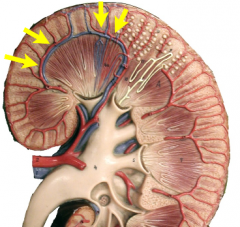
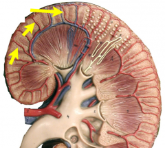
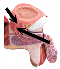
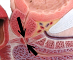
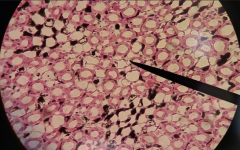
What is this covering? |
Renal capsule |
|
What is this covering? |
Renal capsule |
|
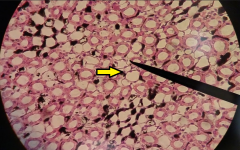
What is this covering? |
Adipose capsule |
|
What is this covering? |
Renal fascia |
|
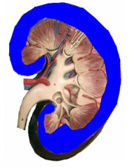
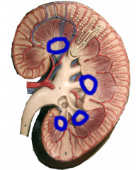
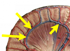
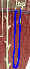
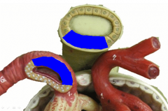
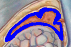
What is this region (highlighted in blue)? |
Renal cortex |
|
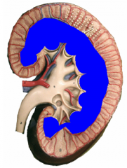
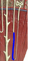
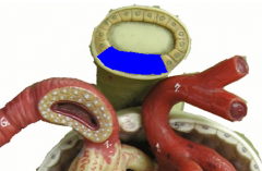
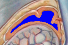
What is this region (highlighted in blue)? |
Renal medulla |
|
What is this region? |
Renal cortex |
|
What is this region? |
Renal medulla |
|
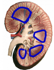
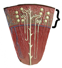
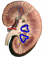
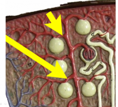
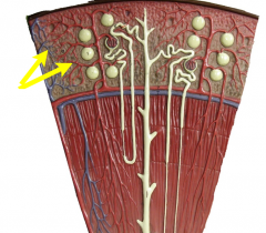
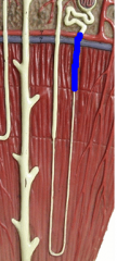
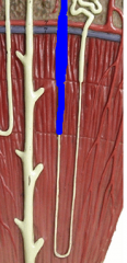
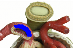
What are these structures (highlighted in blue)? |
Renal pyramids |
|
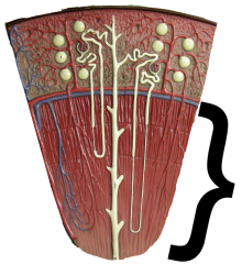
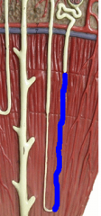
What are these structures (highlighted in blue)? |
Renal pyramids |
|
What are these specific structures (highlighted in blue)? |
Renal papillae (pl.) Renal papilla (sing.) |
|
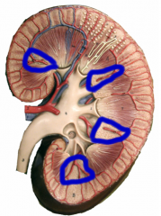
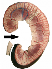
What are these structures (highlighted in blue)? |
Renal columns |
|
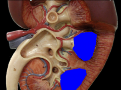
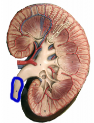
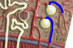
What are these structures (highlighted in blue)? |
Minor calyces (pl.) Minor calyx (sing.) |
|
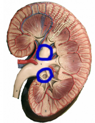
What are these structures (highlighted in blue)? |
Major calyces (pl.) Major calyx (sing.) |
|
What is this structure (highlighted in blue)? |
Major calyx |
|
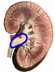
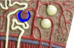
What is this structure (highlighted in blue)? |
Renal pelvis |
|
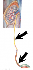
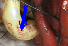
What is this structure (highlighted in blue)? |
Ureter |
|
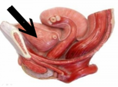
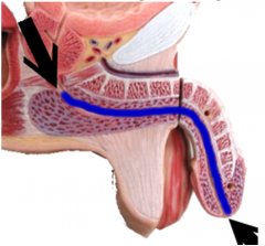
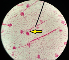
What is this structure? |
Ureter |
|
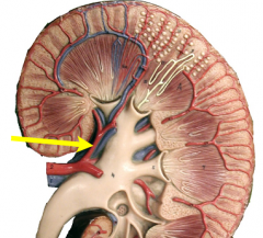
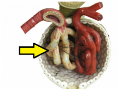
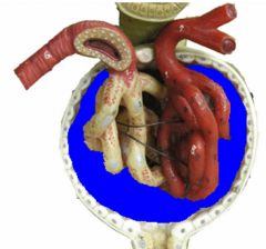
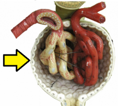
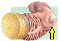
What is this space? |
Renal sinus |
|
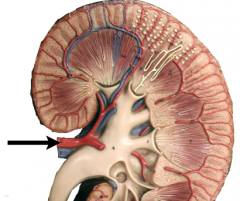
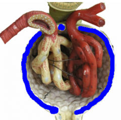
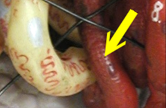
What is this structure? |
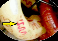
Renal artery |
|
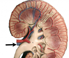
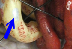
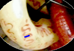
What is this structure? |
Renal vein |
|
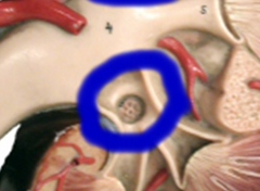
What is the structure at the tip of the pointer? |
Segmental artery |
|
What are the structures at the tips of the pointers? |
Interlobar arteries |
|
What is the structure at the tip of the pointer? |
Interlobal vein
|
|
What are the structures at the tips of the pointers? |
Arcuate arteries |
|
What is the structure at the tips of the pointers? |
Arcuate vein |
|
What are the structures at the tips of the pointers?
|
Interlobular arteries |
|
What is the structure at the tips of the pointers? |
Interlobular arteries |
|
What are the structures at the tips of the pointers? |
Afferent arterioles |
|
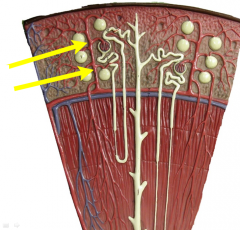
What is the structure at the tip of the pointer? |
Glomerulus |
|
What are these structures (highlighted in blue)? |
Efferent arterioles |
|
What are the structures at the tips of the pointers? |
Peritubular capillaries |
|
What is the structure at the tips of the pointers? |
Vasa recta |
|
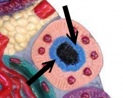
What are the structures at the tips of the pointers (the entire spherical structure)? |
Renal Corpuscles |
|
What is this structure (highlighted in blue)? |
Glomerulus |
|
What is the structure highlighted in blue? |
Bowman's capsule |
|
What specific layer is this (highlighted in blue)? |
Parietal layer of Bowman's capsule |
|
What is the specific layer (in white) at the tip of the pointer? |
Visceral layer of Bowman's capsule (covering glomerulus) |
|
What is this space (highlighted in blue)? |
Bowman's space |
|
What is the entire spherical structure? |
Renal corpuscle |
|
What is the opening at the tip of the pointer? |
Fenestration |
|

What tissue layer is at the tip of the pointer? |
Fenestrated endothelium |
|
What is the cell at the tip of the pointer? |
Podocyte |
|
What is the cell at the tip of the pointer? |
Podocyte |
|
What is the feature at the tip of the pointer? |
Pedicel |
|
What is the space at the tip of the pointer? |
Filtration slit |
|
What is the feature (highlighted in blue)? |
Pedicel |
|
|
What are the 3 stages of urine formation? |
1. Glomerular filtration 2. Tubular reabsorption 3. Tubular secretion |
|
What is the function of the specific layer at the tip of pointer? |
Prevent blood cells & platelets from entering filtrate |
|
What is the specific layer (in blue) at the tip of pointer? |
Basal lamina of Glomerulus |
|

What is the specific layer (in red) at the tip of pointer? |
Basal lamina of Podocytes |
|
What is the function of the specific layer at the tip of pointer? |
Prevent lg. proteins from entering filtrate |
|
What are the specific features at the tips of pointers? |
Pedicels |
|
What are the spaces in between the specific features called? |
Filtration slits |
|

What is the function of the spaces at the tips of the pointers? |
Prevent medium proteins from entering filtrate |
|
What is this structure (highlighted in blue)? |
Proximal convoluted tubule (PCT) |
|
What is this structure (highlighted in blue)? |
Loop of Henle (LOH) |
|
What is the specific structure (highlighted in blue)? |
Thick descending loop of Henle |
|
What is the specific structure (highlighted in blue)? |
Thin descending loop of Henle |
|
What is the specific structure (highlighted in blue)? |
Thin ascending loop of Henle |
|
What is the specific structure (highlighted in blue)? |
Thick ascending loop of Henle |
|
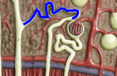
What is this structure (highlighted in blue)? |
Distal convoluted tubule (DCT) |
|
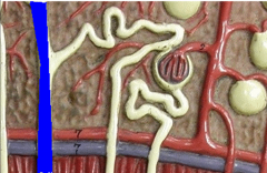
What is this structure (highlighted in blue)? |
Collecting duct (CD) |
|
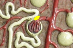
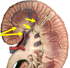
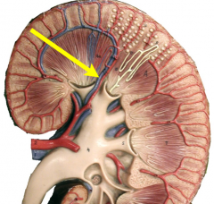
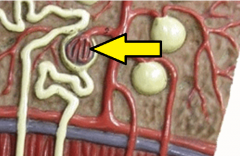
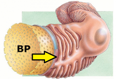
What is this feature (highlighted in yellow)? |
Juxtaglomerular apparatus (JGA) |
|
What is this feature (highlighted in blue)? |
Juxtaglomerular apparatus (JGA) |
|
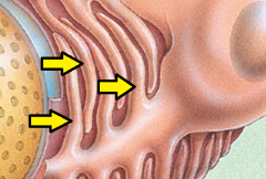
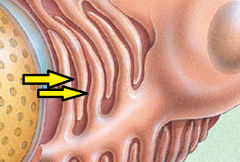
What specific cells are these (highlighted in blue)? |
Macula densa cells |
|
What specific cells are these (highlighted in blue)? |
Juxtaglomerular cells |
|

What are the these structures? |
Proximal convoluted tubules (PCT) |
|
What specific tissue type is found here? |
Simple cuboidal ET |
|
What specific feature (in blue) is shown here? |
Brush border of PCT |
|
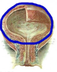
What is this structure (outlined in blue)? |
Urinary bladder |
|
What is this structure (outlined in blue)? |
Urinary bladder |
|
What is this structure (outlined in blue)? |
Urinary bladder |
|
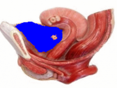
What is this structure (outlined in blue)? |
Urinary bladder |
|
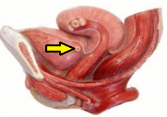
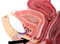
What is this structure at tip of pointer? |
Ureter |
|
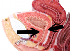
What are these openings at tips of pointers?
|
Orifices of ureters |
|
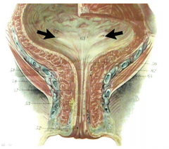
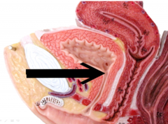
What is this opening (outlined in black)? |
Internal urethral orifice |
|
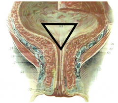
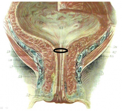
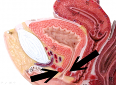
What is this specific region (outlined in black)? |
Trigone |
|
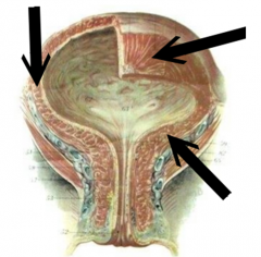
What is this structure (at tips of arrows)? |
Detrusor |
|
What is this structure (at tips of arrows)? |
Detrusor |
|
What structure is this (at tip of arrow)? |
Detrusor |
|

What is this structure at tips of arrows? |
Urethra |
|
What is this circular band of muscles? |
Internal urethral sphincter |
|
What is this opening? |
Internal urethral orifice |
|
What is this opening? |
External urethral orifice |
|
What is this circular band of muscles? |
External urethral sphincter |
|

What is this opening (highlighted in blue)? |
Urethra |
|
What is this specific structure? |
Prostatic urethra |
|
What is this specific structure? |
Membranous urethra |
|
What is this specific structure? |
Cavernous (penile) urethra |
|
What is this opening? |
External urethral orifice |
|
What is this structure? |
Thyroid gland |
|
What are these structures? |
Parathyroid glands |
|
What are these structures? |
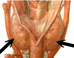
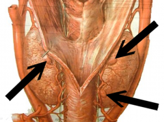
Adrenal glands |
|
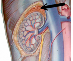
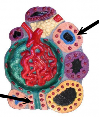
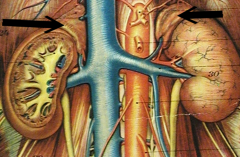
What is this structure? |
Adrenal gland |
|
What is this region (highlighted in blue)? |
Adrenal cortex |
|
What is this region (highlighted in blue)? |
Adrenal medulla |
|
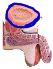
What is this specific specimen? |
Urinary bladder |
|

What state is this organ in? |
Expanded |
|

What is the specific layer at the tip of the pointer? |
Muscularis |
|

What structure is at the tip of the pointer? |
Middle circular layer of muscularis |
|

What is the name of the compilation of structures at tip of pointer? |
Detrusor |
|
Name the layer at the tip of the pointer |
Adipose capsule |
|

What region contains renal corpuscles? |
Renal cortex |
|

What specific region separates the cortex from the medulla? |
Corticomedullary junction |
|

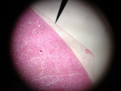
Identify the specific surface indicated by the pointer |
Lateral surface |
|
Identify the structure at the tip of pointer |
Thin limb of loop of Henle (3 simple squamous cells) |
|

Identify the structure to the left of the pointer |
Thick limb of loop of Henle (3 simple cuboidal cells) |
|

Identify the structure to the right of the pointer |
Collecting duct |
|
Identify the structure at the tip of the yellow arrow |
Vasa recta |
|

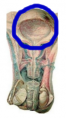
Identify the specimen in the field of view |
Bladder |
|
What state is this organ in? |
Contracted |
|

What is the structure at the tip of the pointer? |
Detrusor muscle |
|

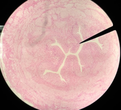
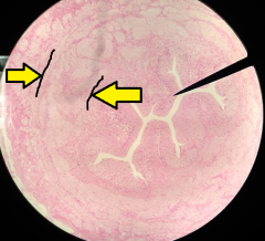
What is the specimen in the field of view? |
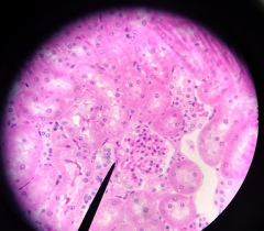
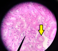
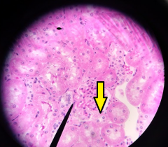
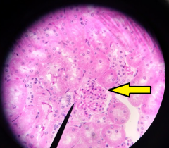
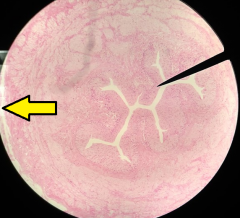
Kidney |
|

What main structure is the pointer pointing to? |
Renal pyramid |
|
What is the cup-like structure the structure at the pointer is draining into? |
Calyx |
|

What is the apex of the structure at the tip of the pointer called? |
Renal papilla |
|

What does the apex of the structure at the tip of the pointer point towards? |
Renal hilum |
|
What is this specific specimen? |
Kidney |
|

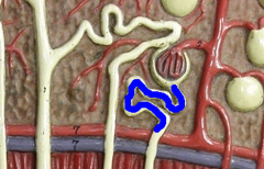
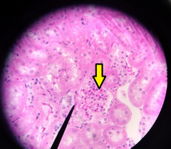
What cell is found at the tip of the pointer? |
Juxtaglomerular (JG) cells |
|
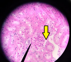
What is the structure at the tip of the yellow arrow? |
Glomerulus |
|
What space is at the tip of the yellow arrow? |
Bowman's space |
|
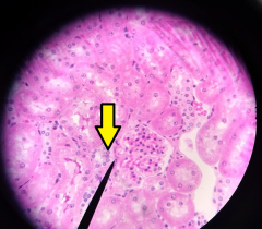
What cells are at the tip of the yellow arrow? |
Macula densa |
|
What is the structure at the tip of the yellow arrow? |
PCT |
|
What is the structure at the tip of the yellow arrow? |
DCT |
|
What layer is at the tip of the yellow arrow? |
(Parietal layer of) Bowman's capsule |
|

What collective structure is at the tip of the pointer? |
Renal corpuscle |
|

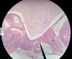
What is the specimen in the field of view? |
Kidney |
|
What is the structure at the tip of the pointer? |
Interlobular artery |
|
What is the structure at the tip of the yellow arrow? |
Afferent arteriole |
|
What are the structures at the tips of the yellow arrows? |
Peritubular capillaries |
|

What hormone regulates the constriction of the structure at the tip of the yellow arrow? |
Angiotensin-II |
|
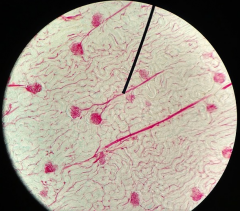
What is the specimen in the field of view? |
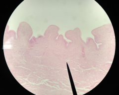
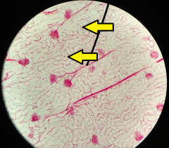
Ureter |
|

What is the specific layer at the tip of the pointer? |
Mucosa |
|

What is the tissue type at the tip of the pointer? |
Transitional epithelial tissue |
|
What is the general layer in between the two yellow arrows? |
Muscularis |
|

What are the specific layers in between the two yellow arrows? |
--Inner longitudinal --Outer circular |
|
What is this layer at the tip of the yellow arrow? |
Adventitia |
|
|
What hormones are secreted by the kidney & what are their functions? |
--Erythropoietin (EPO): stimulates erythropoiesis --Calcitriol: form of vitamin D that ↑blood calcium by promoting absorption of dietary Ca --Renin: ↑BP |
|
|
What are the components of the filtration membrane & what do they do? |
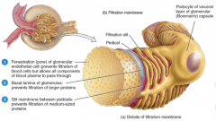
--Fenestrated endothelium: prevents filtration of blood cells & platelets --Basal lamina: prevents filtration of lg. proteins --Slit membrane: prevents filtration of med. proteins |
|
|
What are the pressures that influence GFR & how do they relate to net filtration pressure? |
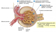
--Glomerular Blood Hydrostatic Pressure (GBHP): 55 mmHg --Capsular Hydrostatic Pressure (CHP): 15 mmHg --Blood Colloid Osmotic Pressure (BCOP): 30 mmHg Net Filtration Pressure = GBHP - CHP - BCOP = 55 mmHg - 15 mmHg - 30 mmHg = 10 mmHg |
|
|
From where do cortical nephrons receive their blood supply? |
Peritubular capillaries |
|
|
From where do juxtamedullary nephrons receive their blood supply? |
Vasa recta |
|
|
What feature differentiates the LOH in cortical nephrons from the LOH in juxtamedullary nephrons? |
Cortical nephrons w/ short LOHs have only thick ascending limbs in their LOH Juxtamedullary nephrons w/ long LOHs have thick & thin ascending limbs in their LOH |
|
|
What is blood hydrostatic pressure? |
Main hydrostatic pressure that pushes water & solutes through filtration membrane, promoting filtration |
|
|
What is capillary hydrostatic pressure? |
Pressure exerted against the filtration membrane by fluid in the capsular space, opposing filtration |
|
|
What is blood osmotic pressure? |
Pressure of plasma proteins "pulling" on water, opposing filtration |
|
|
Why must GFR be regulated? |
Maintain homeostasis |
|
|
What are the mechanisms of GFR regulation? |
--Renal autoregulation: kidneys themselves regulate GFR --Neural regulation: ANS regulates renal blood flow & GFR --Hormonal regulation: angiotensin-II & ANP |
|
|
How much filtrate is received by the PCT? |
80 mL/min |
|
|
How much filtrate is receied by the LOH? |
40 - 45 mL/min |
|
|
What is the primary function of Na+ in the body? |
Fluid-electrolyte balance |
|
|
Where is most of the Na+ in the body found? |
ECF |
|
|
What hormones regulate body Na+ levels? |
--ADH --ANP --Aldosterone |
|
|
What is the primary function of Cl- in the body? |
Maintain anion balance in fluid compartments |
|
|
Where is most of the Cl- in the body found? |
ECF |
|
|
What hormones regulate Cl- levels within the body? |
ADH (follows Na+) |
|
|
What is the primary function of K+ in the body? |
Establishing resting membrane potential |
|
|
Where is most K+ in the body found? |
ICF |
|
|
What hormones regulate K+ levels within the body? |
Aldosterone |
|
|
What is the primary function of HCO3- in the body? |
Buffer |
|
|
What regulates HCO3- levels within the body? |
Intercalated cells |
|
|
What are the primary functions of Ca2+ in the body's fluids? |
--Blood clotting --NT release --Excitability of nervous & muscle tissue |
|
|
What hormone regulates body Ca2+ levels in the body? |
PTH |