![]()
![]()
![]()
Use LEFT and RIGHT arrow keys to navigate between flashcards;
Use UP and DOWN arrow keys to flip the card;
H to show hint;
A reads text to speech;
179 Cards in this Set
- Front
- Back
|

|
|
|
|
|
|
|
|

|

|
|
|
|
|
|

|
|

|
|
|

|

|
|
|

|
|
|

|
|

|

|
|

|

|
|

|

|
|

|

|
|

|

|
|

|

|
|

|

|
|

|

|
|

|

|
|

|
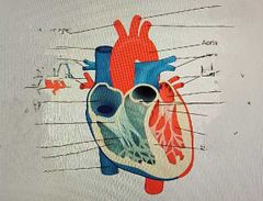
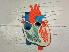
Wiggers Diagram |
|
|
Wiggers Diagram |
A diagram of volumes, pressures, electrical activity, and heart sounds. Used to illustrate the relationship between different aspects of cardiac physiology and the sequence of events in the cardiac cycle |
|
|
P Wave |
Electrical activity that leads atrial contraction |
|
|
A Wave |
Atrial contraction Detected via atrial pressure monitoring |
|
|
QRS Complex |
Electrical activity that leads ventricular contraction |
|
|
S1 |
Heart sound: closure of mitral valve |
|
|
Sharp upward deflection in ventricular pressure |
Ventricular contraction |
|
|
C Wave |
The large pressure increase that occurs in the ventricle with contraction causes the closed tricuspid valve to bulge into the atrial space, raising atrial pressure
Not always seen |
|
|
S2 |
Heart sound: closure of the aortic valve |
|

|
Isovolumic contraction |
|

|
Ejection |
|

|
Isovolumic relaxation |
|

|
Rapid inflow |
|

|
Diastasis |
|

|
Atrial Systole |
|
|
Cardiac Output |
The blood that is ejected from the left ventricle into the aorta over 1 minute |
|
|
Cardiac Output Formula |
CO= HR x SV |
|
|
Cardiac Output Normal Range |
4-8 L/min |
|
|
Cardiac Index |
Compares cardiac output to the size of the body |
|
|
Cardiac Index Formula |
CI= CO/body surface area |
|
|
Cardiac Index Normal Range |
2.5-4 L/min/m^2 |
|
|
Ejection Fraction |
Percentage of the left ventricular end diastolic volume ejected in systole |
|
|
Ejection Fraction Formula |
EF= (SV/end diastolic volume) x 100% |
|
|
Ejection Fraction Normal Range |
60%-80% |
|
|
Systemic Vascular Resistance |
The sum of the forces resisting blood flow through the systemic circulation |
|
|
Systemic Vascular Resistance Formula |
[(mean aortic pressure - right atrial pressure)/CO] x 80 |
|
|
Systemic Vascular Resistance Normal Range |
900-1400 dynes/sec/cm^-5 |
|
|
Pulmonary Vascular Resistance |
The sum of the forces resisting blood flow through the pulmonary circulation |
|
|
Pulmonary Vascular Resistance Formula |
PVR= [(mean pulmonary artery pressure - pulmonary capillary wedge pressure)/cardiac output] x 80 |
|
|
Pulmonary Vascular Resistance Normal Range |
30-100 dynes/sec/cm^2 |
|

|
Aortic valve opens |
|

|
Aortic valve closes |
|

|
Mitral valve closes |
|

|
Mitral valve opens |
|

|
Systole |
|

|
Diastole |
|

|
Systole |
|

|
Phonocardiogram |
|

|
Aortic pressue |
|

|
Ventricular pressure |
|

|
Atrial pressure |
|

|
Ventricular volume |
|

|
Electrocardiogram |
|
|
Mean Right Atrial Pressure Normal Range |
2-8 mmHg |
|
|
Right Ventricular Pressure Normal Range |
Systolic: 15-30 mmHg Diastolic: 1-8 mmHg |
|
|
Pulmonary Artery Pressure Normal Range |
Systolic: 15-30 mmHg Diastolic: 4-12 mmHg |
|
|
Mean Pulmonary Capillary Wedge Pressure Normal Range |
2-10 mmHg |
|
|
Mean Left Atrial Pressure Normal Range |
2-10 mmHg |
|
|
Left Ventricular Pressure Normal Range |
Systolic: 100-140 mmHg Diastolic: 4-12 mmHg |
|
|
Normal value for inferior vena cava O2 saturations |
70 +/-5% |
|
|
Normal value for superior and inferior vena cava O2 saturations |
70 +/-5% |
|
|
Normal value for right atrial O2 saturations |
70 +/-5% |
|
|
Normal value for right ventricular O2 saturations |
70 +/-5% |
|
|
Normal value for pulmonary artery O2 saturations |
70 +/-5% |
|
|
Normal value for left atrial O2 saturations |
95 +/-5% |
|
|
Normal value for left ventricular O2 saturations |
95 +/-5% |
|
|
Normal value for aortic O2 saturations |
95 +/-5% |
|
|
What does a right heart catheterization directly measure? |
Pressures on the right side of the heart and the pulmonary circulation |
|
|
What path does a right heart catheterization take? |
Enters through the femoral, subclavian, internal jugular, or brachiocephalic vein > superior vena cava > R atrium > tricuspid valve > R ventricle > pulmonic valve > pulmonary artery > wedged into a smaller vessel in the pulmonary circulation |
|
|
What indirect measurements can right heart catheterizations be used to collect? |
Pressure on the left side of the heart |
|
|
How can right heart catheterizations be used to indirectly measure left heart pressures? |
Through measurement of a pulmonary capillary wedge pressure, which is indicative of the pressure in the left atrium |
|
|
What is another name for a Swan-Ganz catheter? |
Pulmonary artery catheter |
|

|
Proximal port |
|

|
Port to Thermistor |
|

|
Port to balloon |
|

|
Distal Port |
|

|
Superior Vena Cava |
|

|
Right Atrium |
|

|
Right ventricle |
|
|
Distal Aperture |
The tip of the catheter, where the bulk of measurements are taken. Communicates to the distal port |
|
|
What color is most commonly associated with the distal port? |
Yellow |
|
|
Where is the proximal aperture found on the Swan-Ganz catheter? |
30cm mark |
|
|
What color is most commonly associated with the proximal port? |
Blue |
|
|
What measurements can be collected using the proximal aperture? |
- Central venous pressures or right atrial pressures, depending on the position of the catheter - Cardiac output |
|
|
How is cardiac output measured using the proximal aperture? |
Thermodilution technique |
|
|
What is the thermodilution technique? |
A known volume of injectate (typically 10ml D5W), at a known temperature (room temperature), is injected into the right atrium through the proximal port. Blood temperature is measured at the distal end of the pulmonary artery catheter and injectate temperature is measured as it enters the right atrial lumen. Cardiac output is determined by the change in blood temperature over time |
|
|
What color is most commonly associated with the balloon port? |
Red |
|
|
What purpose does the balloon port serve? |
A syringe is attached for the inflation and deflation of the balloon, which sits at the end of the catheter. Many models include an infusion port for medications, through which vasopressors or other meds may be given |
|
|
Thermistor Port |
An electronic plug that connects to the thermistor |
|
|
What is the function of the thermistor? |
Measuring the temperature change at the distal portion of the catheter during the measurement of cardiac output when using the thermodilution technique |
|
|
Transducer |
The device which converts pressure exerted upon it into an electrical signal, generating the waveforms observed during cardiac catheterization |
|
|
At which point should the transducer be leveled in order to produce accurate readings? |
Phlebostatic axis |
|
|
Phlebostatic Axis |
The intersection of the mid-axillary line and the fourth intercostal space The level of the atrium |
|
|
What equipment be used to ensure accuracy of measurements during cardiac catheterization? |
A leveling device |
|
|
How would the pressure measurements skew if the transducer is sitting below the phlebostatic axis? |
Falsely elevated |
|
|
How would the pressure measurements skew if the transducer is sitting above the phlebostatic axis? |
Falsely lowered |
|
|
Steps to ensure accurate pressure evaluation |
- level and zero transducer - ensure connections are not loose or leaking - remove all bubbles from the circuit - assess the pressure waves and EKG for correlation - Take measurements at the appropriate time in the respiratory cycle |
|
|
V Wave |
Represents right atrial passive filling |
|
|
What does the peak of the V wave represent? |
The opening of the tricuspid valve |
|

|
A wave |
|

|
C wave |
|

|
V wave |
|

Why do the pressure waveforms lag slightly behind the ECG? |
Electrical activity precedes changes in pressure |
|
|
What part of the heart has the lowest pressures? Why? |
Right Atrium It fills passively from venous return |
|

What do the large spikes in pressure above the second picture represent? |
Right ventricular systole |
|

During right ventricular diastole, why does the baseline ventricular pressure return to a pressure equal to the right atrium? |
The tricuspid valve is open during ventricular diastole |
|

|
Dicrotic notch |
|
|
Dicrotic Notch |
The notch in the downslope of the pulmonary artery pressure wave, caused by the closure of the pulmonic valve |
|

Why are the baseline pressures above the final picture higher than those observed in the right atrium and ventricle? |
Pulmonary wedge pressure represents pressures in the left side of the heart, which is a higher pressure area |
|
|
If it is difficult to determine which waveform is the a wave due to HR or clinical status, what could be used as an indication? |
The p wave should be found on the ECG and correlated to the waveform that immediately follows it |
|
|
What is the possible indication of a very large a wave? Why? |
Stenosis of the tricuspid valve The force of the atrial contraction must be stronger than usual to open a stenotic valve |
|
|
How would the a wave appear if the patient is in atrial fibrillation? |
There would be no a wave |
|
|
V Wave |
Occurs at the end of ventricular systole, after the T wave on the ECG Represents left atrial filling against a closed mitral valve |
|
|
How can one use physical assessment to determine which pressure wave is the V wave? |
The V wave will be seen just after the pulse is felt |
|
|
Low right atrial pressures can be indicative of what conditions? |
Dehydration or hypovolemia |
|
|
High right atrial pressures may be indicative of what conditions? |
- Heart failure - Fluid volume overload - Tricuspid valve disease - Atrial septal defect |
|
|
Why must one be cautious when advancing the catheter into the R ventricle? |
As the catheter bumps into the walls of the ventricle, it can cause PVCs/vtach |
|
|
What should one do if vtach occurs during advancement into the ventricle? |
The catheter may need to be pulled back slightly In some cases, cardioversion may be required |
|

|
Contraction of the R ventricle |
|
|
Increased systolic pressures can be seen in what conditions? |
- pulmonic stenosis - pulmonary hypertension |
|
|
At what point is right ventricular end diastolic pressure measured? |
At the end of diastole, just before systole |
|
|
What can right ventricular end diastolic pressure indicate? |
- General health of R ventricle - Ventricular preload |
|
|
In what conditions can right ventricular end diastolic pressure become elevated? |
- Right ventricular myocardial infarction - Untreated pulmonary stenosis - Untreated pulmonary hypertension |
|
|
Is it more difficult to advance the catheter into the R ventricle or pulmonary artery? Why is this? |
Pulmonary artery The pulmonic valve is smaller than the tricuspid valve, so some manipulation may be required |
|
|
When entering the pulmonic artery, what changes can be observed? |
A distinct increase in pressures The disappearance of the atrial kick |
|

What does the upward deflection (#1) on the pulmonary artery waveform represent? |
Right ventricular systole |
|

What does the descent marked #2 on the pulmonary artery waveform represent? |
Right ventricular diastole |
|

What does the second, smaller upward deflection (#3) represent on the pulmonary artery waveform? |
Dicrotic notch Transient pressure increase related to pulmonic valve closure |
|
|
Increased pulmonary artery pressures can be a result of what conditions? |
- COPD - pulmonary hypertension - pulmonary embolus - left-to-right shunting - left sided heart failure - hypoxemia - valve disease |
|
|
How is pulmonary artery wedge pressure obtained? |
The inflated balloon is advanced until it "wedges" in a smaller vessel in the pulmonary circulation. The balloon creates a seal against the walls of the vessel, isolating the tip of the catheter from pressure changes behind the balloon |
|
|
How much air is used to inflate the balloon in a pulmonary artery catheter? |
1.5mL |
|
|
How long can a pulmonary artery catheter be wedged, and why is there a limit? |
No longer than 15 seconds, or about two respiratory cycles The balloon is blocking blood flow through the blood vessel, essentially acting as a pulmonary embolus |
|
|
When should pulmonary wedge pressure be measured, and why does this matter? |
Near the end of expiration Endothoracic pressure fluctuates with breathing, causing wedge pressure measurements to vary. Inhalation causes negative endothoracic pressures |
|
|
How might sedation affect wedge pressure measurements? |
Heavily sedated patients may exhibit long, snoring respiratory patterns. This may make measurements less accurate |
|
|
What might an upward trend in wedge pressure indicate, and what can be done? |
The balloon may be overinflated,or the vessel may be too small. The balloon may need to be deflated and the catheter repositioned |
|
|
What might cause elevated pulmonary wedge pressures? |
- Left ventricular overload - COPD - Mitral regurgitation - Mitral stenosis - Myocardial infarction affecting the left ventricle - Heart failure - Aortic stenosis - Aortic regurgitation |
|

|
A wave Represents left atrial contraction |
|

|
C wave Represents bulging of the mitral valve during ventricular systole |
|

|
Atrial relaxation and the downward movement of the mitral valve |
|

|
V wave Represents passive filling of the left atrium |
|

|
The opening of the mitral valve and ventricular filling |
|
|
What conditions might cause increased left atrial pressures |
- Mitral stenosis - Mitral regurgitation - Left ventricular infarction - CHF - Aortic stenosis - Aortic regurgitation |
|
|
When are left ventricular end diastolic pressures measured? |
Just before systole |
|
|
Under what circumstances are left ventricular end diastolic pressure typically elevated? |
- volume overload - valve disease - diastolic dysfunction - cardiomyopathy |
|
|
What procedure is required to directly measure left heart pressures? |
Left heart catheterization |
|

What is represented by the highlighted area on this right atrial waveform? |

Tricuspid regurgitation
As the ventricle contracts, blood is forced back into the atrium, raising atrial pressure and causing a very large v wave |
|
|
How can one identify mitral regurgitation according to heart pressure measurements? |
A large v wave on a left heart pressure waveform |
|

What is indicated by these waveforms? How can this be determined? |
Mitral stenosis Normally, the left atrial diastolic pressure will be close to the left ventricular end diastolic pressure. In this diagram, the left atrial pressures are high because the stenotic valve is not allowing normal emptying of the left atrium into the left ventricle. This blood, retained in the atrium, results in higher atrial pressures |
|
|
What conditions are associated with valve stenosis? |
- rheumatic fever - drug abuse - advanced age - congenital defects |
|

What is indicated by this diagram? How can this be determined? |
Aortic valve stenosis Normally, the gradient between LV pressures and aortic pressures is very small, with systolic pressure in the ventricle being slightly higher than aortic systolic pressure.
Here, high left ventricular pressures with significantly lower aortic pressures can be seen. This is because the left ventricle must generate a large amount of pressure to force blood through the narrowed valve. The lower aortic systolic pressure is the result of a lower volume of blood entering the aorta |
|

What is represented by this diagram? |
Aortic valve pullback technique, showing aortic valve stenosis |
|
|
Aortic Valve Pullback |
A technique in which the catheter is advanced through the aortic value into the left ventricle, then pulled back into the aorta. LV and AO pressures are measured on two sequential beats, evaluating for pressure differences that would indicate aortic stenosis |
|
|
How are pressures affected by pulmonic valve stenosis? |
Right ventricular systolic pressure is high, with low pulmonary artery systolic pressure |
|
|
How is the severity of pulmonic valve stenosis measured? |
By the width of the gradient between right ventricle systolic pressure and pulmonary artery systolic pressure |
|
|
What blood oxygen saturation abnormalities would you expect in a patient with a left to right shunt through an atrial septal defect? Why? |
The saturations in the right atrium would be much higher than those found in the inferior and superior vena cava This is due to the oxygenated blood crossing from the left atrium to the right atrium through the septal defect |
|
|
Is it possible to have a right to left shunt through a septal defect? Why or why not? |
It is possible but much rarer due to the typically higher pressures on the left side of the heart |
|
|
What is a general indication of a shunt somewhere in systemic circulation? |
A sharp decrease in blood oxygen saturation at any point other than the tissues, or a sharp increase in blood oxygen saturation anywhere other than the lungs |
|
|
What complication can result from a ventricular septal defect? |
Heart failure |
|
|
How is a ventricular septal defect corrected? |
Surgery or cardiac catheterization |
|
|
What abnormal blood oxygen saturation measurements would you expect in a patient who has a left to right shunt through a ventricular septal defect? Why? |
Saturation in the right ventricle that are higher than that in the right atrium
This is due to oxygenated blood flowing from left ventricle into the right ventricle through the septal defect |
|
|
In the case of a right to left shunt through a ventricular septal defect, what blood oxygen saturations would be expected? |
Right atrium and right ventricle would have a similar saturation Left ventricular and aortal saturations would be decreased compared to the left atrium and pulmonary vein |
|
|
Which conditions make up Tetralogy of Fallot? |
Pulmonic stenosis Dextroposition of the aorta (aka overriding aorta) Ventricular septal defect Thickened right ventricle |
|
|
Dextroposition of the Aorta |

A congenital heart defect where the aorta overrides the interventricular septum to the right ventricular side |
|
|
Symptoms of mild Tetralogy of Fallot |
Heart murmur Intermittent cyanosis Squatting |
|
|
Symptoms of severe Tetralogy of Fallot |
Severe, continuous cyanosis heart murmur Boot-shaped appearance of the heart on radiographs Fainting spells ("tet spells) |
|
|
Why do children with Tetralogy of Fallot tend to squat or pull their knees to their chest? |
These positions increase vascular resistance, allowing a transient reversal of a portion of the shunting |
|
|
Ductus Arteriosus |
Shunts blood from the pulmonary artery to the aorta in fetal circulation Typically closes within a week of birth |
|
|
Symptoms of a patent ductus arteriosus |
Depending on the pressures involved, there is either left to right or right to left shunting (most frequently from the aorta into the pulmonary circulation, raising pressures in the pulmonary circulation Hypoxemia SOB Widening of pulse pressure |
|
|
What type of surgical repair is used for patent Ductus Arteriosus |
Ligation of the vessel |
|
|
Pharmacological treatment for patent ductus arteriosus |
Indomethacin Helps constrict the ductus arteriosus by inhibiting prostaglandin E2 synthesis |