Reading...
![]()
Play button
![]()
Play button
![]()
Use LEFT and RIGHT arrow keys to navigate between flashcards;
Use UP and DOWN arrow keys to flip the card;
H to show hint;
A reads text to speech;
34 Cards in this Set
- Front
- Back
|
Simple Squamous epithelium location
|
Lines BV (as endothelium), pleual, peritoneal and serous cavities (mesothelium)
-Bowmans Capsule, thin loop of Henle |
|
|
Simply Cuboidal epithelium Location
|
Lines distal tubules of kidney, follicles of thyroid gland, and surface of ovary
|
|
|
Simple Columnar epithelium location
|
Are polyhedral cells.
Line stomach, intestine and excretory ducts |
|
|
Non-keratinized stratified squamous location
|
Moist body surface, esophagus and vagina
|
|
|
Stratified Cuboidal location
|
Lines duct of sweat glands.
|
|
|
Stratified Columnar location
|
In large excretory ducts and in cavernous urethra.
|
|
|
Pseudo-stratified Epithelium location
|
Trachea, bronchi, ducts of parotid gland, epididymis
|
|
|
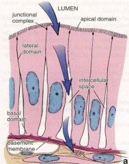
Terminal bar
|
Is composed of the junctional complexes,
ZO, ZA, and MA. |
|
|
Zonula Occludens (tight junctions)
|
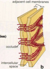
Is the fusion of the outer leaflets. These surround the entire apical perimeter of the cell. Are held together via occluding.
|
|
|
Zonula Adherens
|
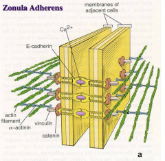
Also extend completely around the perimeter, just basal to the ZO.
Are reinforced by a mat of actin filaments to which E-cadherin, catenin and vinculin anchor to. Central molecules is Ca+. Are intermediate junctions, or belt desmosomes, similar to tight junctions. |
|
|
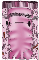
Macula Adherins (desmosomes)
|
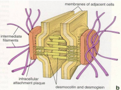
Has dense plaques on the cytoplasmic side. With desmogleins and E-cadherin transmembrane glycoproteins.
There are intermediate (keratin) filaments that loop into and out of the plaques. Are transmembrane links that help stabilize the structure. |
|
|
Connexons (gap junctions)
|
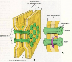
Made up of connexin protein central channels. Also for the passage of ions and small molecules from cell to cell.
In epithelium, CNS, cardiac and smooth muscle. |
|
|
What pathway of fluid movement in intestine?
|
From the lumen into the cell, then into the intercellular space then across the basement membrane to the connective tissue.
|
|
|
What are lateral interdigitations?
|
They are finger like or irregular projections that interlock adjacent epithelial cell.s
|
|
|
Basement membrane (basal lamina)
|
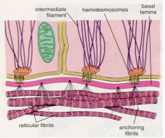
Composed of type IV collagen, laminin and proteoglycans.
Two zones: Lamina Lucida - low density, lies apically Lamina Densa: dense reticular fibril network into which the lamina lucida anchors. |
|
|
Hemidesosomes
|
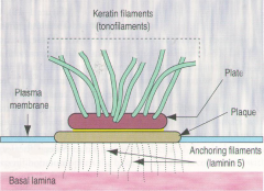
Are incomplete desmosomes (half of a desmosome)
Present at basal surface of cells and anchors the cell into basal lamina. Contains intracellular plaque with keratin filaments loop out. Has transmembrane integrins anchoring into reticular fibers of basal lamina (laminin 5). |
|
|
What is the function of basal plasma membrane foldings?
|
It a common specialization in ion transporting epithelia, from deep invaginations that bring Ion pumps into closer contact with compartmentalized mitochondria to get ATP.
|
|
|
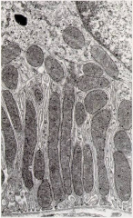
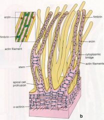
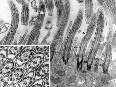
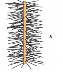
Microvilli
|
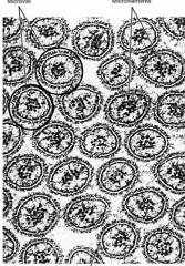
FInger-like projection that increase absorptive surface area, comprise brush border of kidney, and intestinal cells.
Have a glycocalyx (sugar coat). Are a bundle of 30 actin filaments arranged longitudinally that extend into the terminal web of apical cytoplasm. |
|
|
Stereocilia
|
Are long microvilli (not cilia).
Present in epididymis and vas deferens. Also contain bundle of actin filaments that extend into terminal web. |
|
|
Cilia
|
Each clium contains axoneme, core of longitudinal microtubules with 9 doublets (axoneme) surrounding 2 single microtubules.
Has arms in unidirectional fashion with dyenim ATPase, which uses ATP to move cilium. Has a cylindrical basal body at the base of each cilium, which a core of microtubule triplets. |
|
|
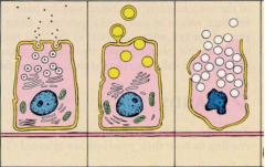
Merocrine
Apocrine Holocrine |
|
|
|
Collagen Location:
Type I Type II Type III Type IV Type V |
I: Bone, tendon, dentin and skin
II: Hyaline and elastic cartilage III: reticular of basement membranes IV: basal lamina V: Aminion and Chorion in fetus |
|
|
Structure of collagen
|
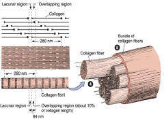
Is a triple a-chain helix containing glycine, hyroxyproline and hydroxylysine.
Gives a transverse banding pattern due to staggered and repeated arrangement of subunits of fibrils. Synthesis: induced by hormones, inhibited by steroids Degraded by MMP. |
|
|
Reticular FIbers:
|
Type III collagen - exhibit banding pattern
Short, fine branches that form scaffold for lymphoid tissue and organs with volume change (BV) Stains well with PAS, exhibit argyrophilia (affinity for silver stain) Secretion by - fibroblasts, smooth muscle cells |
|
|
Elastic Fiber
|
Contains 3 types of fibers: oxytalan, eluanin and elastin
Located: elastic arteries, aorta and branches and vocal folds. Produced: by fibroblasts and smooth muscle cells |
|
|
Ground substance
|
Is the space between cells and fibers
Contains proteglycans, glycosaminoglycans, glycoproteins. |
|
|
Glycosaminoglycans
|
Is a component of ground substance.
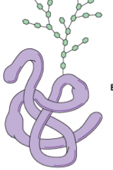
They are repeating disaccharide units composed of a --uronic acid and hexosamine. Are abundant in OH, SO4 and COOH bonds. Form rigid structure to provide for support of cells. Hyaluronic Acid -lack SO4 -large carbohydrate chain -provide barrier for invasion of microorganism Cell surface receptors |
|
|
Proteoglycans
|
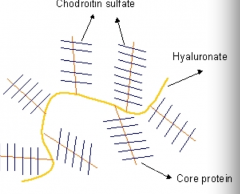
Is a component of ground substance with a protein core to which many GAGs are attached ex, chodrotin sulfate.
Are usually found attached to cell membranes linking matrix to cells. Cell surface recptor |
|
|
Glycoprotein
|
Are globular protein molecules, with branched chain of monosaccharides.
Help stabilize the ECM and links it to the cell surface. |
|
|
Mucous Connective Tissue
|
Is embryonic connective tissue located at umbilical cord.
Called Wharton's jelly. Has few fibers, few cells and abundant ECM with GAGS. Prevents knotting of the cord |
|
|
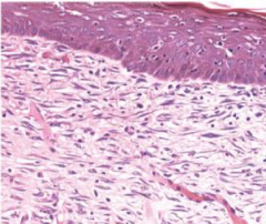
Loose Areolar Connective Tissue:
Type 1 collagen Location: sub-epithelial Has few thin fibers with abundance of migratory cells and ground substance. Vascular with many small blood vessels, nerves, ideal for nutritional support for organs - high metabolic activity |
|
|
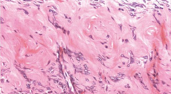
Dense Regular Connective Tissue:
Type I collagen Few cells, little ground substance, more fibers. Location: tendons, ligaments (tensile strength) Collagen fibers are aligned on same axis. |
|
|
Dense Irregular Connective Tissue:
Location: reticular dermis, submucosa Type 1 collagen Few cells, little ground substance and more fibers. The fibers are arranged at random. |
|
|
Brown Adipose Tissue
Predominant in infants, around abdomen and neck. Are multilocular. Dissipate heat. Brown coloration due to abundant deposits of cytochrome oxidase. |