![]()
![]()
![]()
Use LEFT and RIGHT arrow keys to navigate between flashcards;
Use UP and DOWN arrow keys to flip the card;
H to show hint;
A reads text to speech;
16 Cards in this Set
- Front
- Back
|
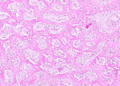
Osteosarcoma
*Mosaic pattern with interspersed osteoid (pink) that is being over produced. *Highly malignant *occurs at age 20 around the knee |
|
|
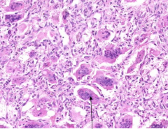
Giant Cell Tumor
*Arrow points to multi-nucleated osteoclast *Benign but locally aggressive *Commonly occur in Epiphysis of any bone at age 20-40 |
|
|
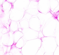
Lipoma
*Most common benign soft tumor in adults *Clear cell appearance w/ small nuclei pushed off to one side |
|
|
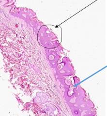
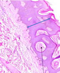
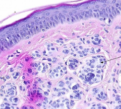
Seborrheic Keratosis
*Circled area shows acanthosis (thickening of epidermis) *Blue arrow points to a horned cyst (aka a keratin pseudocyst) |
|
|
Seborrheic Keratosis
*Black arrow points to a keratin pseudocyst (aka Horned cyst) *Blue shows thick epidermis (acanthosis) |
|
|
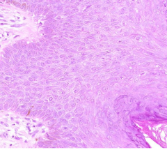
Acanthosis of Seborrheic Keratosis
i.e. thick epidermis |
|
Taken from a raised skin leison |
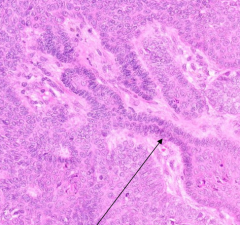
Basal Cell Carcinoma
*Nodules of blue basal cells in dermis (arrow) |
|
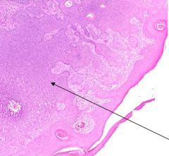
Taken from a raised skin leison |
Basal Cell Carcinoma
*Cells at the periphery of basal tumor nodules in dermis have a palasade "picket-fence" pattern |
|
|
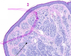
Intradermal Melanocytic Nevus
*Nests of dark nevus cells in dermis (arrow) only and none at the D-E jxn. Intact epidermis |
|
|
Intradermal Melanocytic Nevus
*Black points to sebacous glands *Yellow points to hair follicle |
|
|
Intradermal Melanocytic Nevus
*Nevus cell nests in dermis only with no nests at the D-E jxn. Intact epidermis |
|
|
Bullous Pemphgoid
*Separation of epidermis (red) from dermis (blue) forming a fluid-filled Sub-epidermal bullae *Due to auto-antibody against hemidesmosomes that anchor basalis layer to dermis |
|
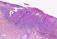
Taken for a 7mm mole |
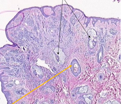
Malignant Melanoma
*Malignant melanocytes are polygonal or spindle shaped and grow in sheets (Black arrow) *cuffed by rim of lighter blue lymphocytes (yellow arrow) *Surface is ulcerated (lighter pink section) |
|
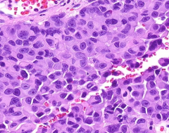
Taken from dermis of a mole sample |
Malignant Melanoma
*Neoplastic melanocytes are polygonal shaped and have large nucleus w/ prominent nucleoli. *Neural Crest derived |
|
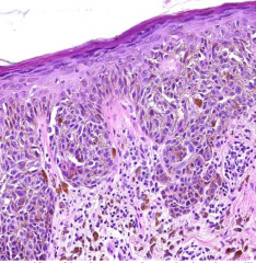
Taken from mole |
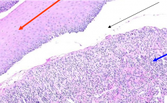
Malignant Melanoma
*Neoplastic cells involving epidermis with extensions into papillary dermis and leakage of melanocytes into dermis
|
|
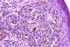
Taken from skin sample |
Malignant Melanoma
*Arrow points to melanin that is being over-produced by neoplastic cells in dermis |