![]()
![]()
![]()
Use LEFT and RIGHT arrow keys to navigate between flashcards;
Use UP and DOWN arrow keys to flip the card;
H to show hint;
A reads text to speech;
73 Cards in this Set
- Front
- Back
|
Cytology |
The study of the microscopic appearance of cells
Used to diagnose diseases by looking at tissue samples from the body |
|
|
Connective Tissue |
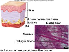
One of the 4 main classes of tissues (epithelial, muscle, and nervous tissues)
Composed of some cells + A LOT of extracellular matrix
Holds things in place
(ex. fascia, tendon, fat, cartilage, bone, skin, and teeth) |
|
|
Extracellular Matrix |
Protein + Carbohydrate mix outside the cell
Provides support to cells
Secreted by the surrounding cells
Different composition in different types of connective tissue |
|
|
Collagen
|
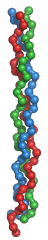
Main structural protein found in extracellular matrix of connective tissue
It is the most abundant protein in mammals
Created by fibroblast cells |
|
|
Fibroblast |
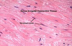
Produces and secretes fibers to maintain the ECM
(ex. collagens, reticular fibers, elastic fibers)
Main cells of connective tissue |
|
|
Epithelium |
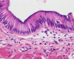
Cells that line a cavity or the outside of the body
Secrete and absorb things
(ex. nutrients from gut, hormones into the blood via endocrine glands in blood, sweat glands on skin) |
|
|
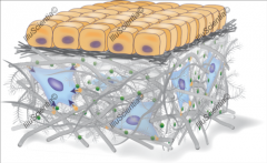
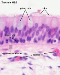
Basal Lamina |
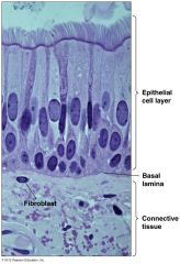
Thin layer of extracellular matrix below epithelial cells
Provides support/protection for Epithelia to sit on (above the muscle, fat, etc)
Composed of proteins/carbohydrates (ex. collagen, proteoglycans, and glycoproteins like laminin and fibronectin)
|
|
|
Reticular lamina
|
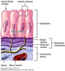
Thin layer of extracellular matrix below the basal lamina
Basal Lamina + Reticular Lamina = "Basement Membrane"
Anchors the basal lamina to underlying connective tissue
Composed mainly of collagen fibers |
|
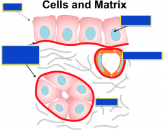
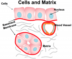
Label:
Cells Nucleus Basement Membrane Blood Vessel Matrix |
|
|
|
2 types of classifications of epithelia based on layers |
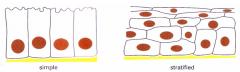
1. Simple (one layer)
2. Stratified (many layers) |
|
|
3 types of epithelium based on cell shape |
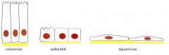
1. Columnar (rectangular)
2. Cuboidal (square)
3. Squamous (flat) |
|
|
Villi |
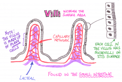
Multiple cells that form "hills" or projections
Increase surface area |
|
|
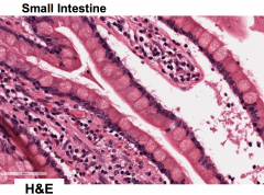
Microvilli |
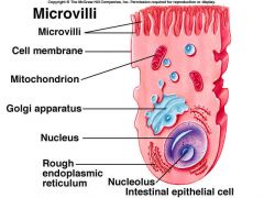
Small little projections from individual cells to increase their surface area
Helps them absorb more substances
(ex. found mainly in the small intestines) |
|
|
Brush Border |
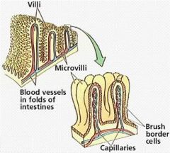
The apical surface of some types of epithelial cells that have microvilli
Specialized for absorption (ex. small intestine and kidney)
Simple cuboidal & simple columnar cells
Enzymes responsible for digestion are tethered as integral membrane proteins in the plasma membrane of the enterocyte. |
|
|
Apical |
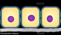
Side of epithelial tissue facing the lumen |
|
|
Basal |
Part of epithelial tissue away from the lumen, touching the basal lamina
(aka basolateral surface) |
|
|
Mucosa |
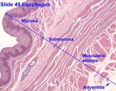
Outer layer of epithelial cells in cavities that are exposed to the external environment (ex. esophagus. nasal passage)
Protects and lubricates |
|
|
Intestinal Gland (aka crypts of Lieberkühn) |
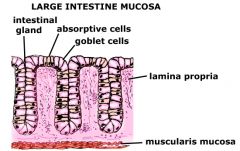
A gland found in the epithelial lining of the small intestine and colon
Inlet of lumen between villi covered by epithelium
Contain stem cells, Paneth cells, and goblet cells |
|
|
Goblet Cell |
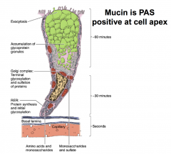
Secretes mucous into the lumen
|
|
|
Paneth Cells |
Found in epithelium of the small intestine
In the crypts of Lieberkühn
Helpful in immune defense (secrete anti-microbial compounds to kill bacteria in the intestinal lumen) |
|
|
Submucosa |
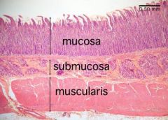
Loose connective tissue directly under a mucous membrane
Supports the mucosa and connects the mucosa to smooth muscle |
|
|
Muscularis externa (aka muscular layer) |
Smooth muscle below the submucosa |
|
|
Adventitia (aka tunica adventitia) |
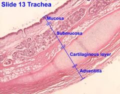
Outermost connective tissue of an organ or structure
|
|
|
Enterocytes |
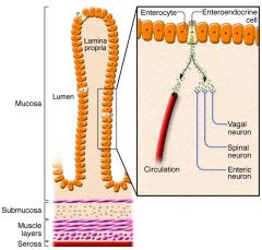
A type of epithelial cell that lines the gut |
|
|
Glycocalyx |
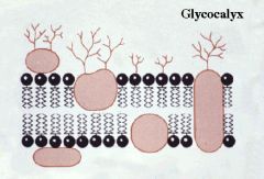
Coat on the outside of cell that is made of carbohydrates
(aka "thin candy shell", like M&Ms)
Found mainly on epithelia & some bacteria |
|
|
Cilia |
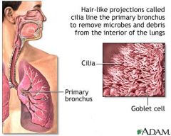
Hairlike fibers on the outside of cell
Made of microtubules
Used to move things along a cavity |
|
|
Fallopian Tube |
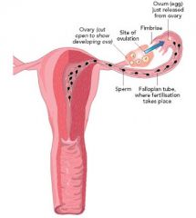
Tubes that carry the egg from the ovary to the uterus
This is where fertilization takes place between a sperm and an ovum
Have cilia |
|
|
Parotid Duct |
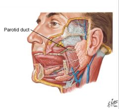
Transports saliva from the parotid gland (aka the major salivary gland) to the mouth
|
|
|
Stages of Mitosis |
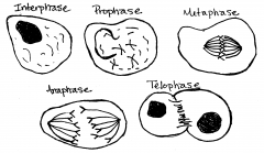
Interphase Prophase Metaphase Anaphase Telophase |
|
|
Hematoxylin |
+ charge (basic)
Stains things BLUE that are basophilic (have + charge)
|
|
|
Eosin |
- charge (acidic)
Stains things RED that are acidophilic (have - charge)
|
|
|
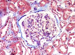
PAS (Periodic acid–Schiff) |
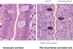
Used to detect carbohydrates (purple color)
(Ex. glycogen, glycoproteins, basement membrane GAGS)
Creates aldehydes from OH groups
|
|
|
Trichrome Stain |
A mixture of 3 dyes used to distinguish different tissue from each other
(ex. collagen fibers stained blue, nuclei stained black, smooth muscle stained red) |
|
|
Silver Stain |
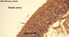
Allows visualization of types of different structures
Specifically for nervous system and reticulin fibers (connective tissue fibers made of type III collagen)
Target tissue appears as black dots |
|
|
Van Gieson Stain |
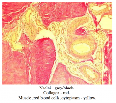
Shows contrast between collagen and other types of tissues
(ex. collagen stains pink, muscle stains yellow) |
|
|
Black “India” Ink Stain |
Used to paint the edges of tumors to see if there are any malignant cells remaining
Used during surgery |
|
|
Iron Hematoxylin Stain |
Used to identify bacteria / parasitic organisms
Also used to visualize mitochondria |
|
|
Steps in tissue preparation |
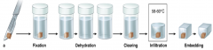
1. Fixation 2. Dehydration (with ethanol) 3. Clearing 4. Infiltration 5. Embedding (in parafin solution) |
|
|
Fixation |
Putting tissue in preservatives
Cross-linking proteins prevents degradation
|
|
|
Parafin |
Oily, petroleum based solution |
|
|
Microtome |
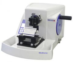
Cuts samples thin (to 5 microns) |
|
|
3 main parts of a light microscope |
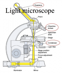
Condenser – collects and focuses light
Objective – enlarges and projects image to eyepiece
Eyepiece - further magnification and projection onto retina |
|
|
How does immunochemistry work to visualize cell components? |
Antibodies recognize proteins in the sections |
|
|
When is EM used? |
Used in renal pathology to diagnose disease |
|
|
Tight Junctions |
Where the membranes of two cells come very close together and appear to be fused.
Create polarity (two different sides of cells), prevent redistribution of the plasma membrane
Act as a barrier so that materials cannot pass between two cells (materials must be regulated by going through the cell)
(ex. in the gut to regulate absorption of digested nutrients)
|
|
|
Three main transmembrane protein families found in tight junctions |
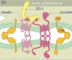
1. Occludin proteins - 2 intracellular and 2 extracellular domains, involved with the regulation of signaling event
2. Claudins proteins - major structural and functional elements, 2 intracellular and 2 extracellular domains, mediate calcium-independent cell-cell adhesion
3. JAMs - single transmembrane domain |
|
|
Adherens Junctions |
Provide strong mechanical attachments between adjacent cells
(ex. hold cardiac muscle cells tightly together, hold epithelial cells together)
|
|
|
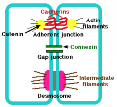
Main types of proteins involved in Adherens Junctions |
cadherins - transmembrane proteins, extracellular segments bind to each other
Only create adhesion when bound to calcium ion
catenins - connect cadherins to actin filaments
Inherited mutations in a gene encoding a cadherin can cause stomach cancer. |
|
|
Nectins |
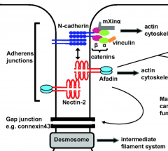
Proteins involved in linking cells to each other extracellularly (like cadherins)
Part of Adherins Junctions |
|
|
Desmosomes |
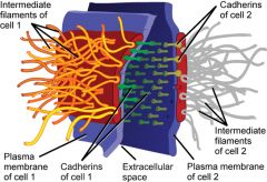
Localized patches that hold two cells tightly together
Appear as thickened patches in the cell membrane region between two cells
Cadherins attach to cytoplasmic plaques that are attached to intermediate filaments (ex. keratin) |
|
|
Hemidesmosome |
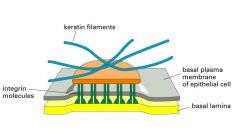
Similar to desmosomes but attach epithelial cells to the basal lamina instead of to each other.
Intracellular plaque is attached to integrins |
|
|
Integrins |
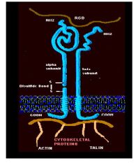
Integrins - mediate cell-ECM interactions with collagen, fibrinogen, fibronectin, and vitronectin.
Provide essential links between the extracellular environment and the intracellular signalling pathways
Play roles in cell behaviours such as apoptosis, differentiation, survival, and transcription
Consist of an alpha and beta subunit, each have a large extracellular domain, a transmembrane domain and a short cytoplasmic domain
|
|
|
Gap Junctions |
Intercellular channels that permit the free passage between of ions and small molecules between cells
Constructed from connexins.
Permit changes in membrane potential to pass from cell to cell (ex. the action potential in cardiac muscle flows from cell to cell through the heart providing the rhythmic contraction of the heartbeat) |
|
|
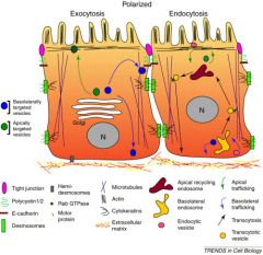
3 Cellular Mechanisms that Create Polarity |
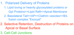
1. Delivery of Proteins (to apical, basal, and lateral areas)
2. Selective Retention, Destruction of Proteins on Apical or Basal Surface
3. Cell-Cell Junctions |
|
|
2 ways to get proteins to the apical membrane |
1. GPI-linked transport (golgi to apical) 2. Transcytosis (basal to apical) |
|
|
GPI Linkage |
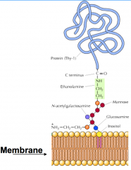
AA sequence on the carboxyl end of a protein that is the signal to send it to the APICAL membrane
These proteins lack transmembrane domains, so they must be associated with lipid or other membrane domains
|
|
|
Lipid Raft |
Takes GPI-linked proteins to the apical membrane
Made of cholesterol and glycosphingolipids
The GpI link is added by recognition of the terminal 30 amino acids of the protein
This stretch of amino acids is “dominant”, and if it is attached to any protein it will send it to the cell surface. |
|
|
Transcytosis |
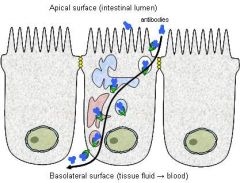
Bringing proteins from the basal to the apical membrane
Ex. immune system antibodies
Mechanism is unclear, but probably doesn't involve a lipid raft |
|
|
Vacuolar Apical Compartment |
A pre-apical compartment that is a stop for golgi=>apical and basolateral=>apical before they reach the apical membrane
This compartment is a bit controversial but it has been seen as a mechanism to quickly collect and load membranes on the apical surface of the cell |
|
|
Hydrophobic XX - tyrosine |
Sequence on a protein to tell it to go to the basal membrane
They stick out of golgi vesicles and bind to adaptor proteins (multi domain, MU domain binds to the XXY region & Beta domain binds to clathrin coating protein)
Clustered in vesicles surrounded by a coat of adaptor proteins and associated clathrin.
|
|
|
Exocyst |
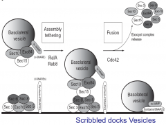
A cluster of nearly 10 proteins which attaches the vesicle to the basolateral membrane.
The vesicle goest to the BL membrane, uncoats its clathrin, and interacts with the exocyst
This permits v-snare and t-snare to attach and vesicle to fuse so the protein gets put into the basolateral membrane
|
|
|
Selective Retention of Proteins |
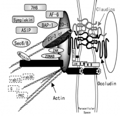
When two cells touch (called a kiss "kiss"), the membrane zone where they meet is very different than the free areas.
When claudins or occluding from different cells are touching, this connection is then built upon by additional claudins and occludins in the interacting cells and they form a plaque of claudins and occludins.
This zone recruits more proteins to aggregate at the site of contact within each cell
(ex. claudin and occluding zone = cytoplasmic plaques of Zo-1, Zonab, cyclins and cytoskeleton)
Adhesion junctions of cadherins also create homotypic patches on the cell membrane (ex. beta-catenin-alpha-catenin-actin filaments organized at those spots) |
|
|
Selective Destruction of Proteins (ex. polymeric IgG receptor called PIGR) |
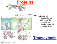
Polymeric IgG receptor = a membrane protein at the basal side of the membrane that binds antibodies (being made by lymphocytes that underlie our epithelial layers)
These antibodies give us what is called “mucosal immunity”
The PIGR binds these antibodies and transcytoses them to the apical lumen, where it is cleaved
This means that full length PIGR is found only at the basal surface and hence appears to be apically distributed. |
|
|
Tight Junctional Compartmentalization |
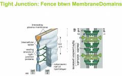
The tight junction forms a “fence” so that the proteins in the lipid membrane cannot undergo lateral movement
The claudins and the occludins block the flow of membrane proteins across their high density zone
Apical remains apical, and basal remains basal |
|
|
Par regulation |
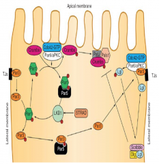
Pars are the regulators of the proportion of the membrane that is apical to the portion of the membrane which is basalolateral.
Par-1 gets phosphorylated by Par-6 and unites with "helper" Par-5
Phosphorylated Par-1along with "helper" Par -5 go to basal space and free Par-3 from the basal part of the tight junction (by Par-1 phosphorylating it too)
Phosphorylated Par-3 then goes with "helper" Par-5 up to the apical side of the tight junction
Hence stimulation of Par1 reduces basolateral Par3 and hence blocks basolateral phenotype, permitting an apical phenotype
|
|
|
Tight Junction Regulation of Cell Proliferation (Cyclins) |
Tight Junctions contain cyclins (proteins that regulate cell proliferation in the nucleus) & Zonab (a carrier protein)
When cells bind via tight junctions, they bind the cyclin in the cell, which diminishes the amount of cyclins entering the nucleus
This reduces cell proliferation
When you have a wound, however, tight junctions are broken and the cyclins engage cell proliferation by being released from the tight junctional plaques so they can enter the nucleus |
|
|
Adhesion Junctions Regulation of Gene Expression (beta-catenin) |
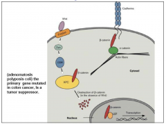
Adhesion junctions contain a reservoir of beta-catenin
When this protein is released into the cytosol it is degraded by APC
If APC is inhibited by Wnt signaling, then excess beta catenin heads into the nucleus and turns on a 1000 genes
This event is the most important signaling in developing stem cells |
|
|
What is absorbed by Epithelial cells? |
Proteins by endocytosis of vesicles coming from the apical membrane
Ions/Water across carriers or channels (simple or facilitated diffusion)
Ions by ATPase |
|
|
What is secreted by Epithelial cells? |
Proteins by exocytosis of vesicles coming from golgi or basolateral face (via transcytosis)
Ions/Water across carriers or channels (simple or facilitated diffusion)
Ions by ATPase |
|
|
4 Rules for Epithelial excretion and absorption |
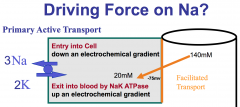
1. Most Carriers-Transporters use energy generated by the NaKATPase (which is on the Basolateral side)
2. Flux is unidirectional (cannot reverse direction)
3. Transport across apical and basal membranes requires different proteins
4. Carriers-Transporters must be correctly positioned (or they will not function)
|
|
|
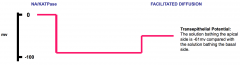
Importance of electrogenic/electroneutral transport |
Electrogenic transport - take into account voltage and concentration gradient
Electroneutral transport - only take into account concentration gradient |
|

Equation to figure out concentration gradient in terms of voltage |
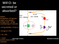
61 log (inside/outside)
Then all you do is compare this to the voltage gradient and use reason to figure out which way each push is sending them
Add or subtract accordingly |
|
|
Membrane potential of apical side vs. basal side |
Apical side is much more negative than the basal side, but still more positive than the cell |